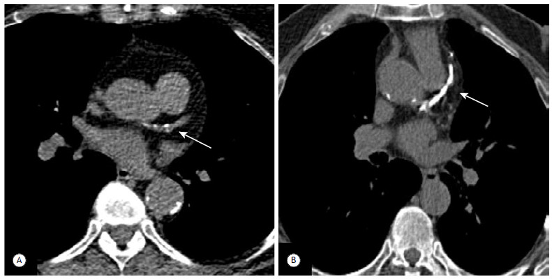
Figure 8. Coronary artery calcifications. In A, axial low-dose CT image showing mild scattered calcified plaques in the proximal left anterior descending coronary artery, findings that should be reported as mild calcifications of the coronary arteries (arrow). In B, severe calcifications of the coronary arteries with heavily calcified plaques distributed along the proximal and mid left anterior descending coronary artery, findings that should be reported as severe calcifications of the coronary arteries (arrow).

