Figure 4.
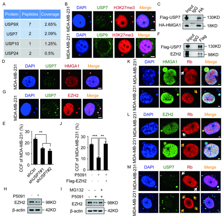
USP7 and HMGA1 stabilize CCF by stabilizing EZH2. (A) Representative results of Flag-HMGA1 combined with mass spectrometry in HEK-293T cells. (B) USP7, H3K27me3, and USP9X were immunofluorescent stained in MM-231 cells to observe the co-localization of USP7 and USP9X with CCF. (C,F) After 48 h of transient transfection of HMGA1, EZH2 and USP7 overexpression constructs in HEK-293T cells, the HA/Flag antibody was used for co-IP. Western blotting was used to detect the interaction between HMGA1 and EZH2 with USP7. (D,G) USP7, HMGA1, EZH2 were immunofluorescent stained in MM-231 cells to observe the co-localization of HMGA1 and EZH2 with USP7. The ratio of CCF was calculated by immunofluorescence in MM-231-shCtrl/MM-231-shUSP7 (E), restoring EZH2 levels of MM-231 cells (J). Western blotting was used to detect the EZH2 level in MM-231 cells treated with P5091 (10 μM) for 48 h (H), MM-231 cells treated with MG132 (10 μM) for 12 h (I). (K–M) MM-231 cells were immunofluorescent stained with HMGA1 and Rb, EZH2 and Rb, and USP7 and Rb to detect the presence of HMGA1, EZH2, and USP7 before (white arrow) or after (yellow arrow) the rupture of the micronuclear membrane. Scale bars = 2 μm. Each experiment was repeated at least 3 times. Error bars, mean ± SD. **, p < 0.01; ***, p < 0.001.

