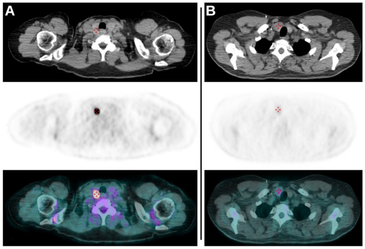
Figure 1.
(A): Axial CT, axial PET and axial fused PET/CT images demonstrating the presence of TI revealed as intense focal uptake of 18F-FDG on the right lobe of thyroid. The lesion had a SUVmax of 44.47, an MTV of 0.7 and a TLG of 18.1 and subsequent cytological exam revealed no malignancy (TIR2). (B): Axial CT, axial PET and axial fused PET/CT images of another scan demonstrating again the presence of TI as a faint uptake on the right lobe of thyroid. The values of SUVmax, MTV and TLG of the lesion were 2.64, 6.9 and 10.3, respectively. Cytological evaluation (TIR5) and subsequent total thyroidectomy revealed the presence of papillary carcinoma.

