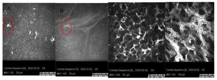
Figure 4.
Images of confocal microscopy of patients with sterile corneal infiltrate after cross-linking procedure. (A) In deep layers of the epithelium, numerous LG cells of varied maturity (circle) are visible. (B) Single inflammatory cells (circle) visible in the layer of Bowman’s membrane with reduced SNP plexus. (C) Site of ulceration with stimulated keratocytes forming a characteristic honeycomblike network. (D) Hyperreflective tissue surrounding keratocytes corresponding to fibrosis.

