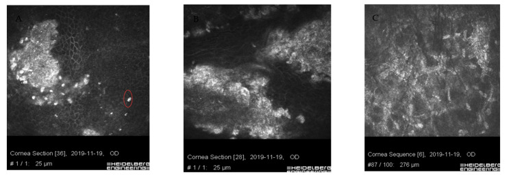
Figure 6.
Images of confocal microscopy of patients with sterile corneal infiltrate after cross-linking procedure. Residual form of keratitis (A,B) with hyperreflective tissue corresponding to scar tissue, irregularly structured epithelial cells with impacted apoptotic hyperreflective cells (circle). (C) Corneal stroma layer with hyperreflective structures corresponding to fibrosis and normal keratocyte nuclei.

