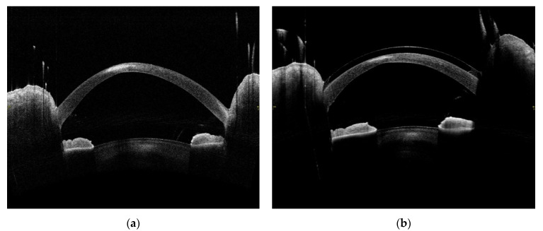
Figure 7.
Images created by the use of optical computed tomography of the anterior segment of the eye of a 20-year-old male patient with peripheral corneal infiltrate after the CXL procedure. (a) Hyperreflective corneal infiltrate seven days after the procedure, 1.83 mm deep and 1.392 mm wide. (b) Hyperreflective residual scar in the corneal stroma six months after the procedure, 0.092 mm deep and 1.323 mm wide.

