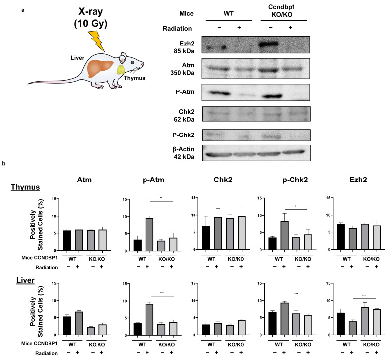
Figure 5.
Effect of Ccndbp1 on the changes in various proteins in X-ray irradiated mice. (a) Western blotting of proteins related with the ATM–CHK2 pathway in mice thymic tissue harvested 12 h after X-ray irradiation. (b) Quantitative analyses of the immunohistochemical analyses of proteins in the thymus and liver of wild (WT) and Ccndbp1 knockout (KO) mice with or without X-ray irradiation. Representative images and a quantitative analysis of positively stained area are shown. The scale bar represents 100 µm. The values represent mean ± standard deviations (n = 5–6), * p < 0.05, ** p < 0.01, *** p < 0.001 on one-way analysis of variance followed by Bonferroni’s multiple comparison test.

