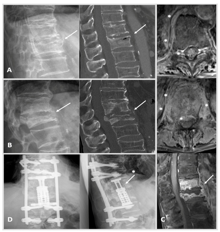
Figure 2.
Case 2: A 66-year-old male patient was treated due to a worsening health status and proven bacteremia (staphylococcus epidermidis and multiresistant enterococcus faecium) of unknown origin. A thoracic CT showed a secondary, suspectedly older, fracture of Th 12 type AO Spine A3 (image (A): left: X-ray; right: CT scan with intravertebral vacuum phenomenon; arrow marking Th 12). MRI imaging was initiated 6 weeks after the image (A) when yet another thoracic CT showed progressing destruction and spondylodiscitis of Th 12 was diagnosed. Initially, CRP was measured at 30 mg/L, and creatinine was documented at 0.99 mg/dL. HSAS risk score: low risk. The patient requested a conservative treatment and agreed to spine surgical transpedicular biopsy, which confirmed the previously identified pathogen S. epidermidis. As primary infectious foci, the patient’s port-catheter was identified and removed promptly, which was in place for a previous esophageal cancer. The patient received anti-infective treatment and returned for follow-up at our out-patient clinic ten weeks later. Here, CRP rose to 100 mg/L, and the patient reported worsening backpain. MRI and CT scans were conducted as well as X-rays standing up showing massive bony destruction of Th 12 and also Th 11 with kyphosis. Image (B): left, X-ray with progressing kyphosis when standing upright; right, CT scan showing pathological fracturing of Th 12 (arrow) type AO Spine A4. Image (C): MRI from top to bottom: free intraspinal conditions cranially with Th 10 surrounding prevertebral tissue reaction marked with stars (axial plane, T1 weighted); more distinctive prevertebral and epidural tissue reaction marked with stars (Th 12 axial plane, T1 weighted); large prevertebral abscess Th10–L1 (arrow) with strong enrichment of Th 11 and Th12 (T1 weighted). Image (D): in a two-step procedure (1) dorsal cement-augmented percutaneous stabilization of Th9–L2 and (2) a lateral minimal invasive thoracotomy approach with incomplete resection of costa 10 was used to achieve anterior stabilization via partial vertebrectomy Th 11 and 12 with debridement and vertebral body replacement with concomitant placement of autologous bone stock i.e., rib (image (D): arrow with star) counteracting kyphosis was established (postoperative X-rays: left anterior-posterior, right lateral view). The patient received anti-infective treatment, and after a long hospitalization, he was able to return to his nursing home. Last follow-up showing after 12 months showed stable clinical findings.

