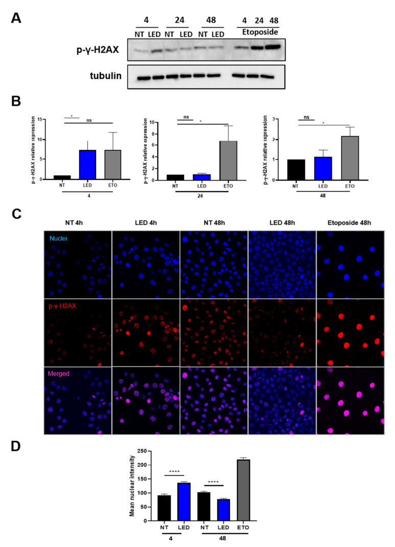
Figure 3.
Exposure to b-LED causes a transient induction of γ -H2AX histone in HaCaT cells. (A) WB showing the expression of γ-H2AX histone (phospho S139) in HaCaT cells exposed to b-LED for 4 h and then analyzed at the indicated times. Etoposide (10 µM) was used as positive control. Western blot analysis was performed on total lysates. Tubulin was detected as a loading control. (B) Densitometric quantification of the experiment shown above. Results are expressed in terms of fold change. (C) Confocal immunofluorescence showing the expression of γ-H2AX histone (phospho S139) in cells stimulated as in (A). (D) quantification of the experiment shown in (C). At least 120 nuclei were quantified from 2 independent experiments. Bars represent the mean ± SEM of duplicate determinations in four independent experiments. * p < 0.05, **** p < 0.0001 and ns, not significant.

