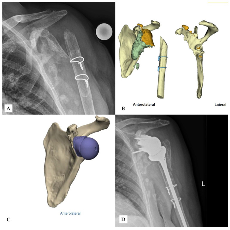
Figure 4.
Second case of revision rTSA. The preoperative X-ray (A) after removal of the prior implant shows extensive damage of the glenoid and the proximal humerus. Bone fragments (orange) and excess cement (grey) can be seen in the CT-scan (B), while the preoperative model (C) shows the scapula after the removal of these fragments. The final outcome shows the glenoid implant in the correct position (D). Reprinted with permission from Materialise. ©2021 Materialise NV.

