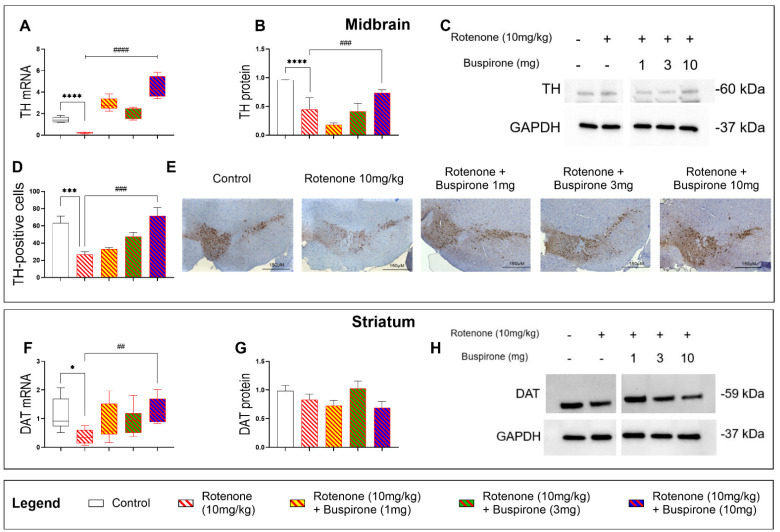
Figure 2.
Buspirone at high dosages protects nigro-striatal dopaminergic neurons from degeneration. TH mRNA (A) and protein expression in the midbrain (B,C). Representative photomicrographs depicting TH immunoreactivity and semi-quantitative measurement of staining intensity in the SNpc, scale bar = 150 µM (D,E). Striatal DAT mRNA (F) and protein expression (G,H). Gene expression was measured by qRT-PCR and quantified using the ΔΔCt method after normalization to s18 (the housekeeping gene). qRT-PCR results are reported as mean fold changes with respect to vehicle-treated control mice. Protein expression was determined by Western blot and normalized to GAPDH (the loading control). Western blots are cropped and removed lanes depicting drug-treatment controls are included in Supplementary Figure S1. Densitometry results are presented as means ± S.E.M. TH-positive cells are reported as the percent of TH-positive cells normalized to the total number of nuclei ± S.E.M. Data represents 3–4 mice per group. * p < 0.05, *** p < 0.001, or **** p < 0.0001, ## p < 0.01, ### p < 0.001, or #### p < 0.0001, as determined by an ANOVA followed by Dunnett’s post-hoc test. TH, tyrosine hydroxylase; DAT, dopamine transporter; GAPDH, glyceraldehyde 3-phosphate dehydrogenase; kDa, Kilodalton; s18, ribosomal protein s18 gene.

