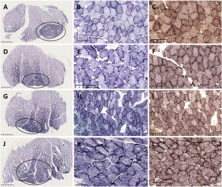
Figure 6.

Soleus muscle pathology in young and ageing wild‐type and Draggen mice. Young wild‐type mouse (A–C). Young Draggen mouse (D–F). Ageing wild‐type mouse (G–I). Ageing Draggen mouse (J–L). Sections stained with NADH‐TR (A,B,D,E,G,H,J,K) and COX‐SDH (G,F,I,L). Scanning magnification view (A,D,G,J) in all animals shows a transverse section through the mid‐belly of the gastrocnemius–soleus muscle. The smaller oxidative, Type I fibre predominant soleus (circle) is present under the larger mixed‐fibre‐type gastrocnemius. In the young wild‐type (B,C) and young Draggen (E,F), the muscle architecture is normal. In the aged wild‐type mouse (H,I), there is subtle uneven oxidative staining in a proportion of fibres, but no overt pathology. In contrast, in the aged Draggen mouse (K,L), both show florid core pathology affecting several fibres, ranging from marked unevenness of oxidative staining, multicores, discrete small cores and occasionally well‐defined larger cores (K,L, arrow). Scale bar: A, D, G, J = 1 mm; N = 50 μm; B, C, E, F, H, I, K, L = 100 μm.
