. 2022 Feb 11;11:e70684. doi: 10.7554/eLife.70684

Figure 4. Growth, ATP, and redox balance of L. plantarum changes when an anode is provided as an extracellular electron acceptor.

These measurements and the current density plot shown in Figure 1C are from the same experiment. (A) Viable cells and (B) dry weight at the point of maximum current density under current circulating conditions (EET) and at open circuit conditions (OC) at the same time point. (C) Change in cell numbers measured by OD600 over time in the bioreactors under EET (continuous line) and OC conditions (dotted lines). (D) ATP production per OD600 unit and (E) NAD+/NADH ratios at the point of maximum current density. The bioreactors were shaken vigorously to dislodge cells before sampling. The avg ± stdev of three biological replicates is shown. Significant differences were determined by one-way ANOVA with (A and B) Dunn-Sidak post-hoc test (n = 3) and (D and E) Dunnett’s post-hoc test (n = 3), * p < 0.05; ** p < 0.01; *** p < 0.001; **** p < 0.0001. See also Figure 1 panel C and Figure 4—figure supplement 3 and related data in Figure 4—source data 1.

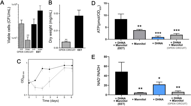
Figure 4—source data 1. Raw data of Figure 4A–D.

Figure 4.

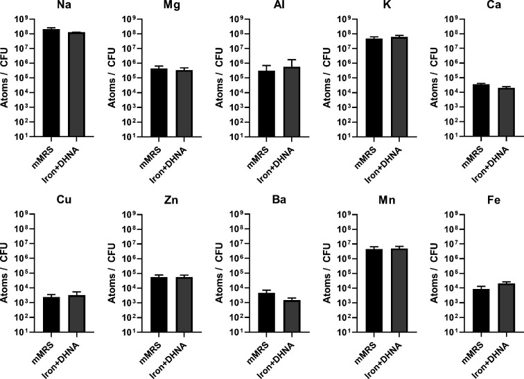
Figure 4—figure supplement 1. Intracellular metal concentrations in L. plantarum are not affected by EET-conducive growth conditions.

Figure 4—figure supplement 1.

Inductively coupled plasma mass spectrometry (ICP-MS) quantification of intracellular metals in L. plantarum after growth for 18 hr in mMRS or mMRS supplemented with 20 μg/mL DHNA and iron (1.25 mM ferric ammonium citrate). The avg± stdev of three biological replicates is shown. See related data in Figure 4—figure supplement 1—source data 1.
Figure 4—figure supplement 1—source data 1. Raw data of Figure 4—figure supplement 1.
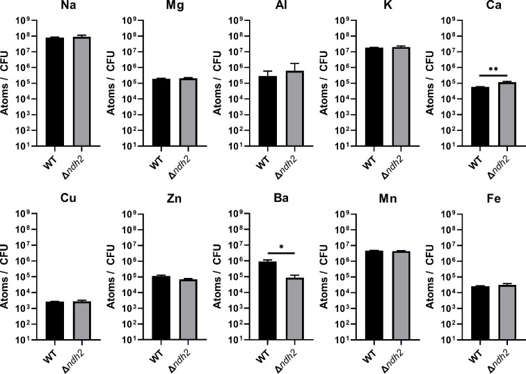
Figure 4—figure supplement 2. Redox-active metal concentrations in L. plantarum are not affected by the presence of ndh2.

Figure 4—figure supplement 2.

Inductively coupled plasma mass spectrometry (ICP-MS) quantification of intracellular metals in wild-type L. plantarum and a L. plantarum ndh2 deletion mutant after growth for 18 hr in mMRS supplemented with 20 μg/mL DHNA and iron (1.25 mM ferric ammonium citrate). The avg ± stdev of three biological replicates is shown. Significant differences determined by one-way ANOVA with Dunnett’s post-hoc test, * p < 0.05; ** p < 0.01. See related data in Figure 4—figure supplement 2—source data 1.
Figure 4—figure supplement 2—source data 1. Raw data of Figure 4—figure supplement 1.
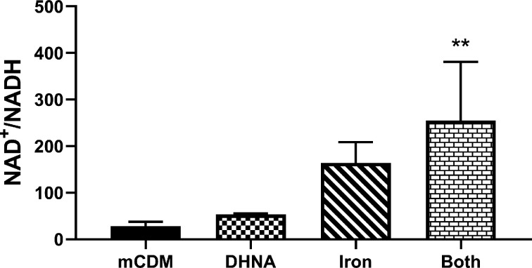
Figure 4—figure supplement 3. Use of Fe3+ as an electron acceptor allows L. plantarum to regenerate NAD+.

Figure 4—figure supplement 3.

NAD+/NADH ratios of L. plantarum grown to mid-exponential phase in mCDM with/without the supplementation of 20 μg/mL DHNA, iron (1.25 mM ferric ammonium citrate), or both. The avg± stdev of three biological replicates is shown. Significant differences determined through one-way ANOVA with Dunnett’s post-hoc test (n = 3), ** p < 0.01. See related data in Figure 4—figure supplement 3—source data 1.
Figure 4—figure supplement 3—source data 1. Raw data of Figure 4—figure supplement 3.