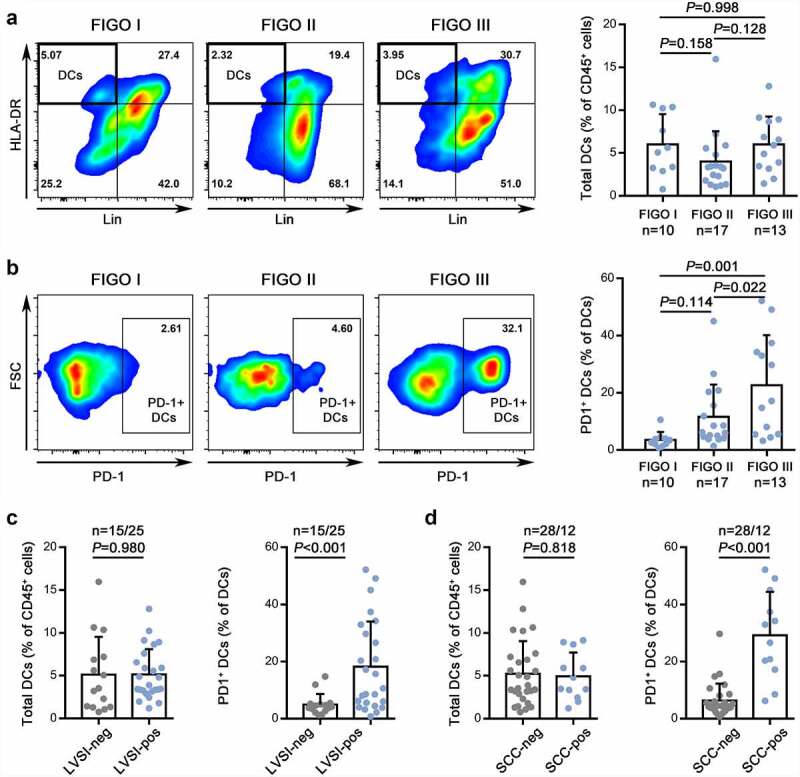
Figure 1.

Identification of enriched intratumoral PD-1+ DCs in advanced CC.
(a,b) Left: The gating strategies for DCs (A) and PD-1+ DCs (B) in CC at different stages. Right: The percentages of DCs (A) and PD-1+ DCs (B) quantified in FIGO stages I, II, and III of CC.(c,d) The percentage of DCs (left) and PD-1+ DCs (right) in CC with or without lymph-vascular space invasion (C) or elevated preoperative SCC antigen levels (D).
