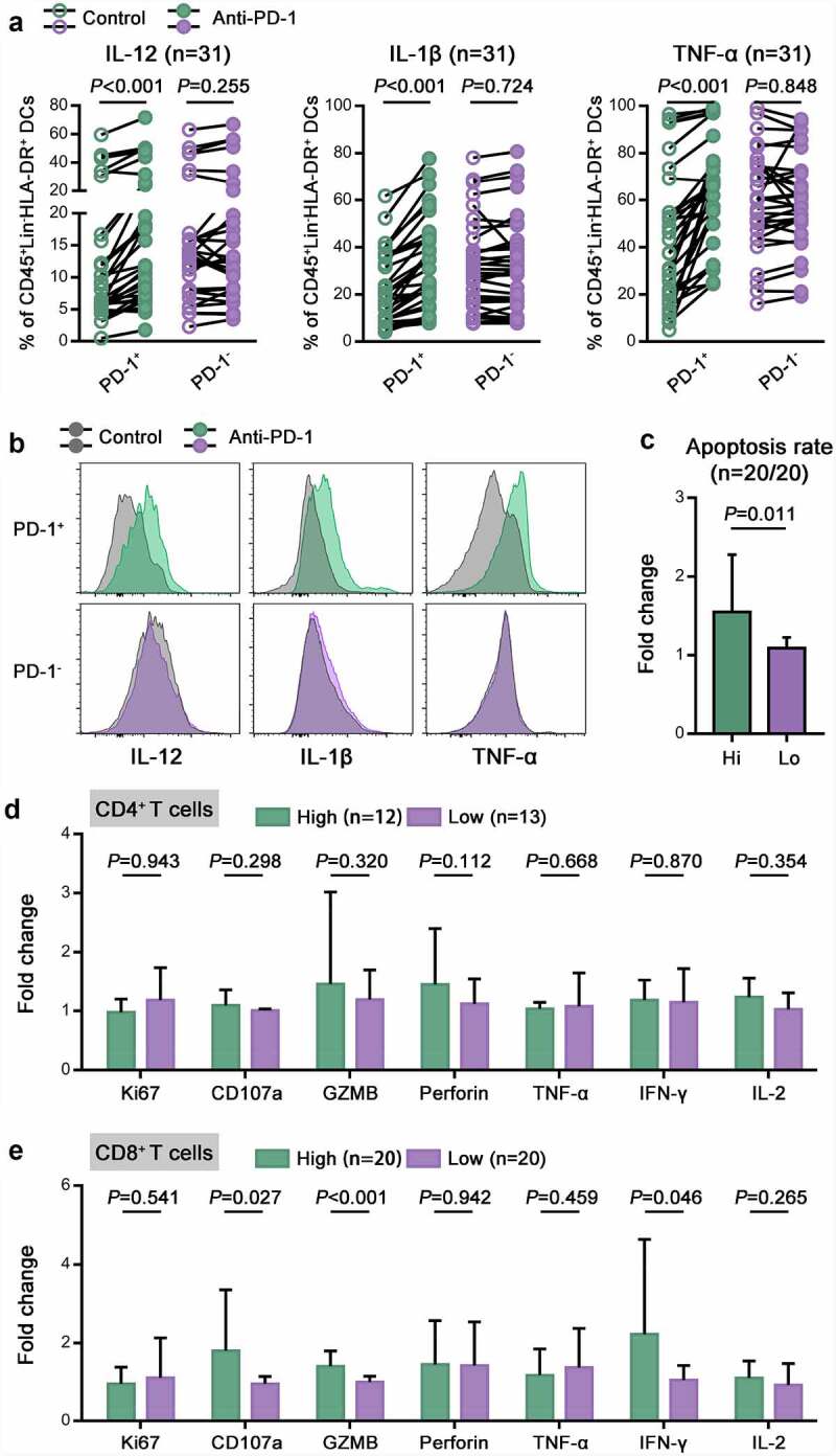
Figure 5.

The accumulation of intratumoral PD-1+ DCs was correlated with a favorable response to PD-1 blockade.
(a) The percentages of IL-12+, IL-1β+ and TNF-α+ cells in PD-1+ or PD-1- DCs from CC tumors before and after treatment with pembrolizumab. (b) Representative flow cytometric histograms showing IL-12, IL-1β and TNF-α expression on PD-1+ or PD-1- DCs in CC before and after treatment with pembrolizumab. (c) The apoptosis rate of tumor cells from high and low PD-1+ DC expressers after treatment, as determined by Annexin V-PI staining. The fold change was calculated as the ratio between the pembrolizumab group and the control group. (d,e) The percentages of Ki67+, CD107a+, GZMB+, perforin+, TNF-α+, IFN-γ+ and IL-2+ cells in CD4 + T cells (D) or CD8 + T cells (E) from high and low PD-1+ DC expressers. The fold change was calculated as the ratio between the pembrolizumab group and the control group.
