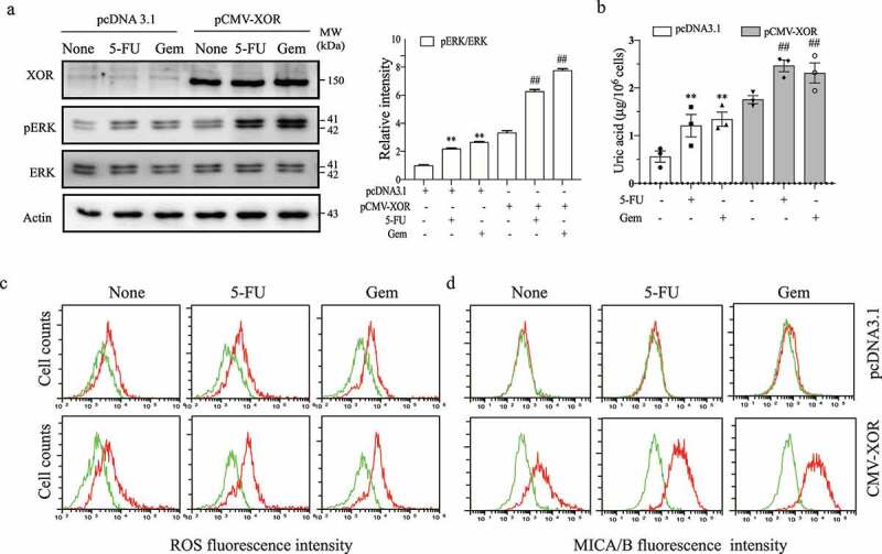
Figure 1.

XOR overexpression enhances genotoxic drug-induced MAP kinase activation, ROS production, uric acid production, and MICA/B expression. MCF-7 cells were transiently transfected with the pcDNA3.1 vector or pCMV/XOR. After incubation for 24 hr, the cells were left untreated or treated with 5-FU (10 μM) or gemcitabine (2 μM) for 24 hr. Cell lysates were prepared and analyzed for ERK phosphorylation by Western blot (a) and for uric acid concentrations by using a uric acid assay kit (b). Relative phosphorylation levels were analyzed by quantifying the density of phosphorylated ERK bands normalized by the density of their total protein bands with NIH Image-J software and presented in a bar graph. Data are the mean ± standard deviation (SD) of three experiments. **p < .01, compared to the untreated control. ##p < .01, compared to the corresponding pcDNA3.1-transfected controls. Gem, gemcitabine. For assaying the levels of ROS and MICA/B, MCF-7 cells were treated as above and incubated for 4 or 24 hr, respectively. Single-cell suspensions were prepared and pulsed with the DCFH-DA fluorescent dye (c) or stained with a PE-conjugated anti-MICA/B antibody (d), followed by analysis with flow cytometry. (c) Green line, unstained; Red line, ROS; (d) Green line, isotype control; Red line, MICA/B. The data represent one of three independent experiments with similar results.
