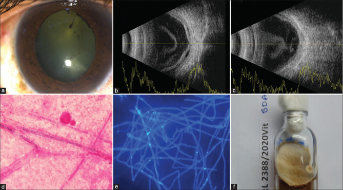
Figure 3.
Case 14, Table 1: A 59-year-old man presented with mild conjunctival congestion (a) and hand motion vision in the right eye. Fundus detail was not visible. Ultrasonogram (USG) of the right eye showed echodense vitreous cavity (b), exudative retina detachment (RD), (b), and choroidal thickening (CT); (c). The vitreous microscopy sample showed septate fungal filaments in direct microscopy [Gram stain, ×1000 (d); Calcofluor white, ×400 (e)]. Fusarium equiseti grew on all media, including Sabouraud dextrose agar (SDA), (f). Treatment included two vitreous procedures (vitrectomy vitreous lavage with silicone oil injection) and 2 times intravitreal amphotericin-B/voriconazole injections. His blood and urine cultures were negative for any organism. At the last follow-up visit (90 days), the eye was quiet; there was extensive scarring of the retina with hand motion vision in the right eye

