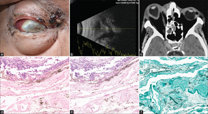
Figure 5.
Case 13, Table 1: A 69-year-old man presented with periocular swelling, discharging fistula, and exudates externally (a) with light perception vision in the right eye. USG showed disorganized eyeball (b), the computer tomography (CT) scan revealed protrusion of the right eye with elongated axial length (c). His eviscerated material and tissue from paranasal sinuses were suggestive of mucormycosis. He received intravenous amphotericin-B and posaconazole. Eyeball was not salvageable; evisceration was done. The histopathology of the eviscerated contents showed broad aseptate fungal filaments with right angle branching suggestive of mucormycosis [hematoxylin and eosin (H and E) stain, ×200 (d); periodic acid Schiff stain (PAS), ×200 (e); Gomori methenamine silver stain (GMS), ×200 (f)]. At 60 days, he expired due to COVID-19-related complications

