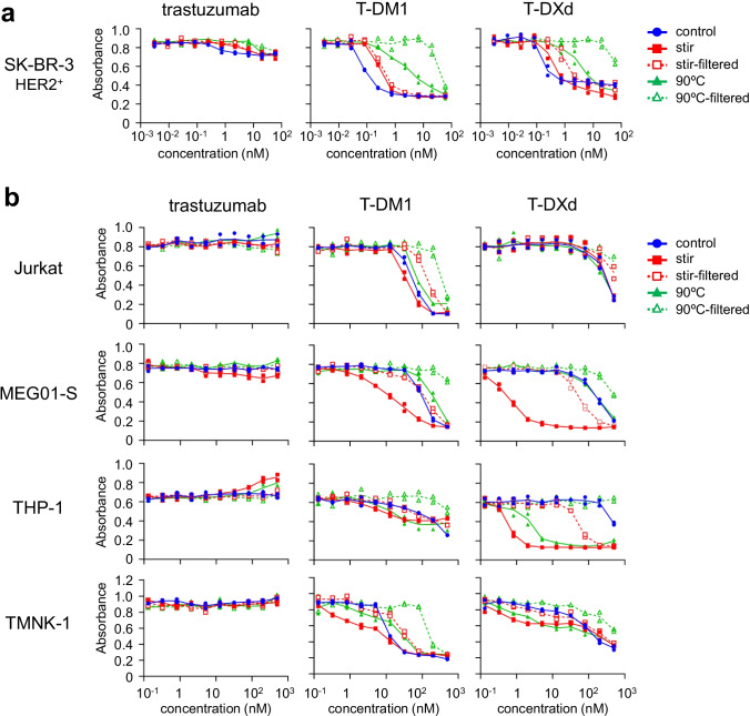
Fig. 2.
The cytotoxicity of ADC aggregates. (a) The cytotoxicity of aggregated ADCs in HER2-positive cells. (b) The cytotoxicity of aggregated ADCs in HER2-negative cells. The HER2-positive cells (SK-BR-3) or HER2-negative cells (Jurkat, MEG-01S, THP-1, TMNK-1) were incubated with serially diluted samples (control, aggregates, or filtrated samples) for 3 days, and the cell proliferations were measured by WST-8 assay. The concentrations of filtered samples represent pre-filtration concentration of the samples. The data represent individual plots (n = 2).

