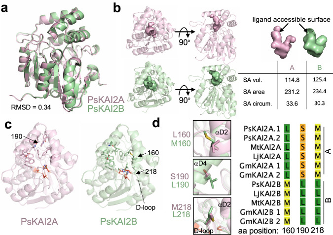
Fig. 7. Structural divergence analysis of legume KAI2A and KAI2B.
a Structural alignment of PsKAI2A and PsKAI2B shown in pink and light green colors respectively. RMSD of aligned structures is shown. b Analysis of PsKAI2A and PsKAI2B pocket volume, area, and morphology is shown by solvent accessible surface presentation. Pocket size values were calculated via the CASTp server. c Residues involved in defining ligand-binding pocket are shown on each structure as sticks. Catalytic triad is shown in red. d Residues L/M160, S/L190, and M/L218 are highlighted as divergent legume KAI2 residues, conserved among all legume KAI2A or KAI2B sequences as shown in reduced Multiple Sequence Alignment from Supplementary Fig. 3.

