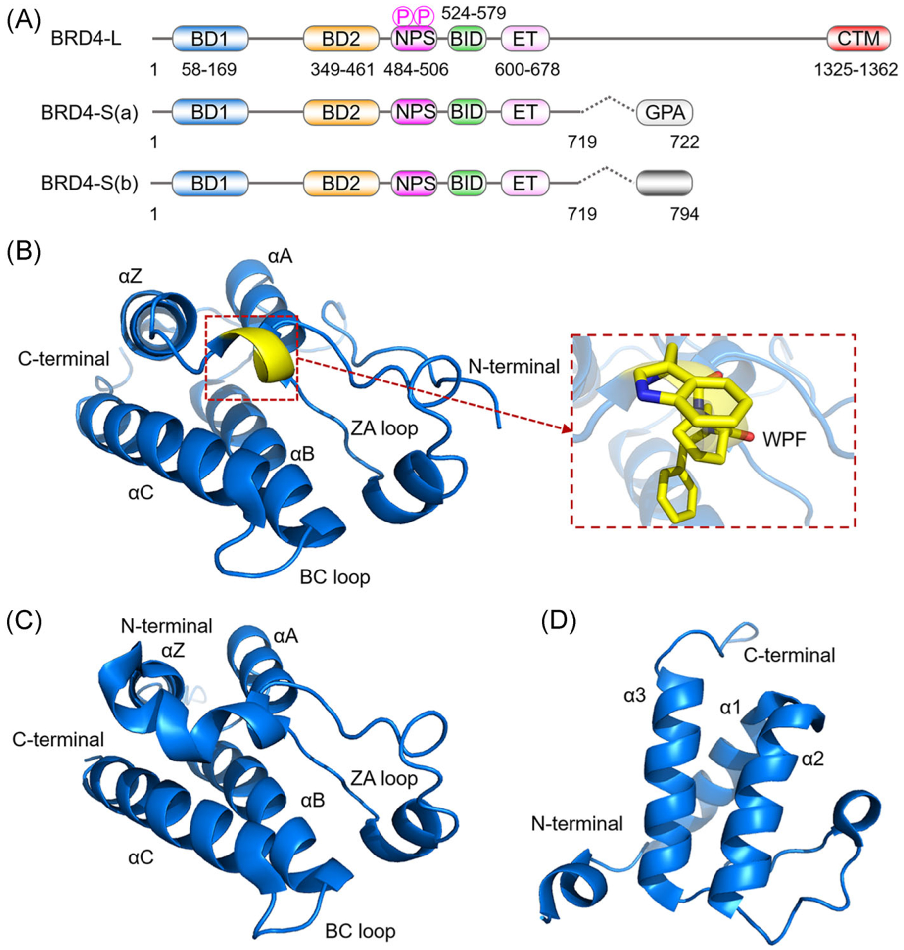
FIGURE 1.

Schematic diagram of BRD4 domain features and crystal structures. (A) Domain architecture of three BRD4 protein isoforms. (B) Crystal structure of BRD4(1) (PDB ID: 3MXF). (C) Crystal structure of BRD4(2) (PDB ID: 5U2C). (D) NMR structure of the BRD4 ET domain (PDB ID: 6BNH). Protein is represented by blue ribbons with WPF shelf residues indicated by yellow sticks
