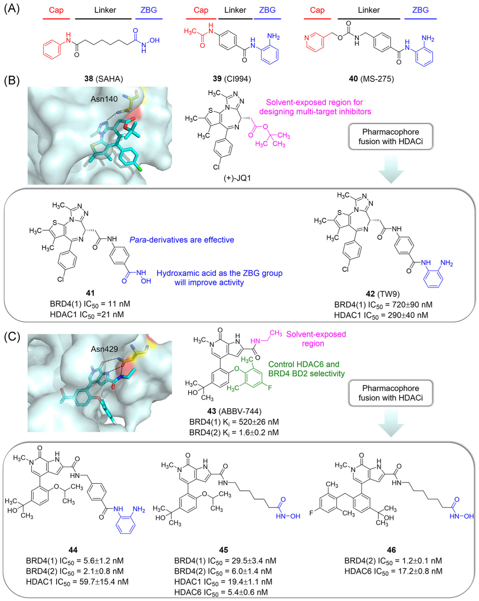
FIGURE 11.

Design of dual-target inhibitors of BET and HDAC based on a pharmacophore fusion strategy. (A) The pharmacophore model of HDACi. (B) Design of compounds 41 and 42. X ray cocrystal structure of JQ1 bound to BRD4(1) (PDB ID: 3MXF). (C) Design of compounds 44, 45, and 46. X ray cocrystal structure of ABBV-744 bound to BRD2(2) (PDB ID: 6E6J). Protein is represented by light blue surface, with compound-contacting residues indicated by yellow sticks. The compound is shown in sticks and colored according to the atom type (C, light blue; N, blue; O, red; Cl, green; F, light cyan). Black dashed line = hydrogen bond
