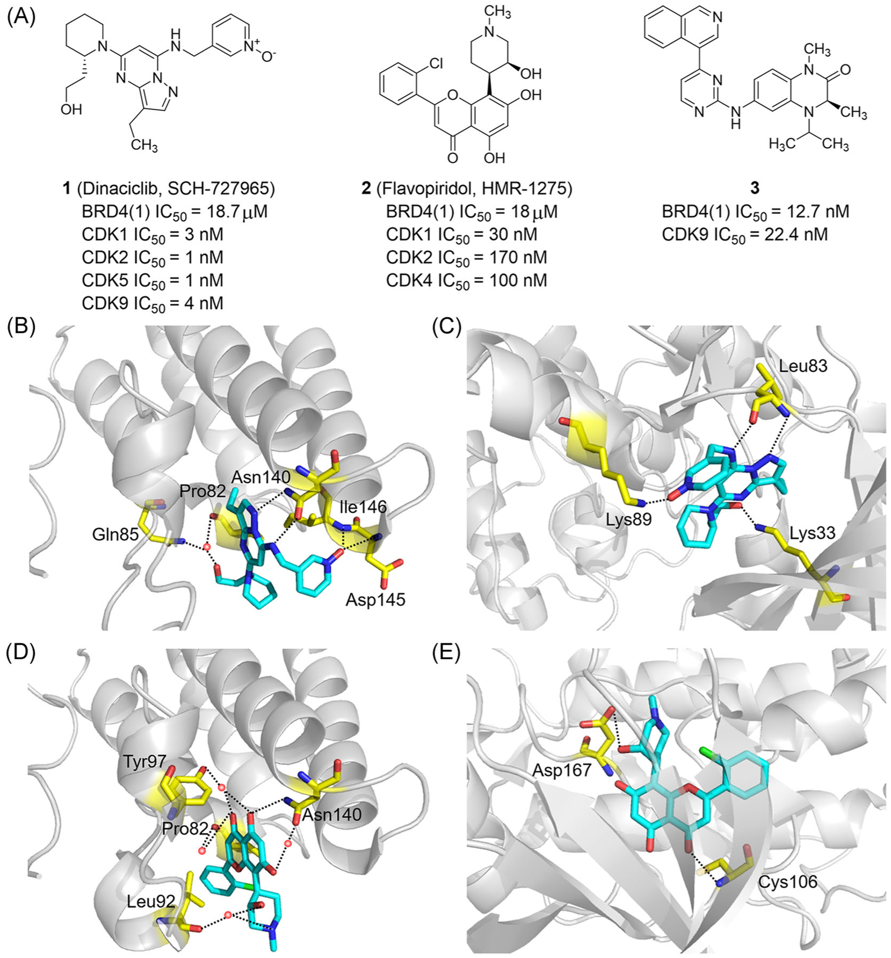
FIGURE 3.

Dual-target inhibitors of BET bromodomains and CDKs. (A) Chemical structures of dinaciclib, flavopiridol and compound 3. (B) X ray cocrystal structure of Dinaciclib bound to BRD4(1) (PDB ID: 4O70). (C) X ray cocrystal structure of dinaciclib bound to CDK2 (PDB ID: 4KD1). (D) X ray cocrystal structure of Flavopiridol bound to BRD4 (PDB ID: 4O71). (E) X ray cocrystal structure of flavopiridol bound to CDK9/cyclin T1 (PDB ID: 3BLR). Protein is represented by gray ribbons, with key compound-contacting residues indicated by yellow sticks. The compound is shown in sticks and colored according to the atom type (C, light blue; N, blue; O, red; Cl, green). Black dashed line = hydrogen bond; orange sphere = water molecule
