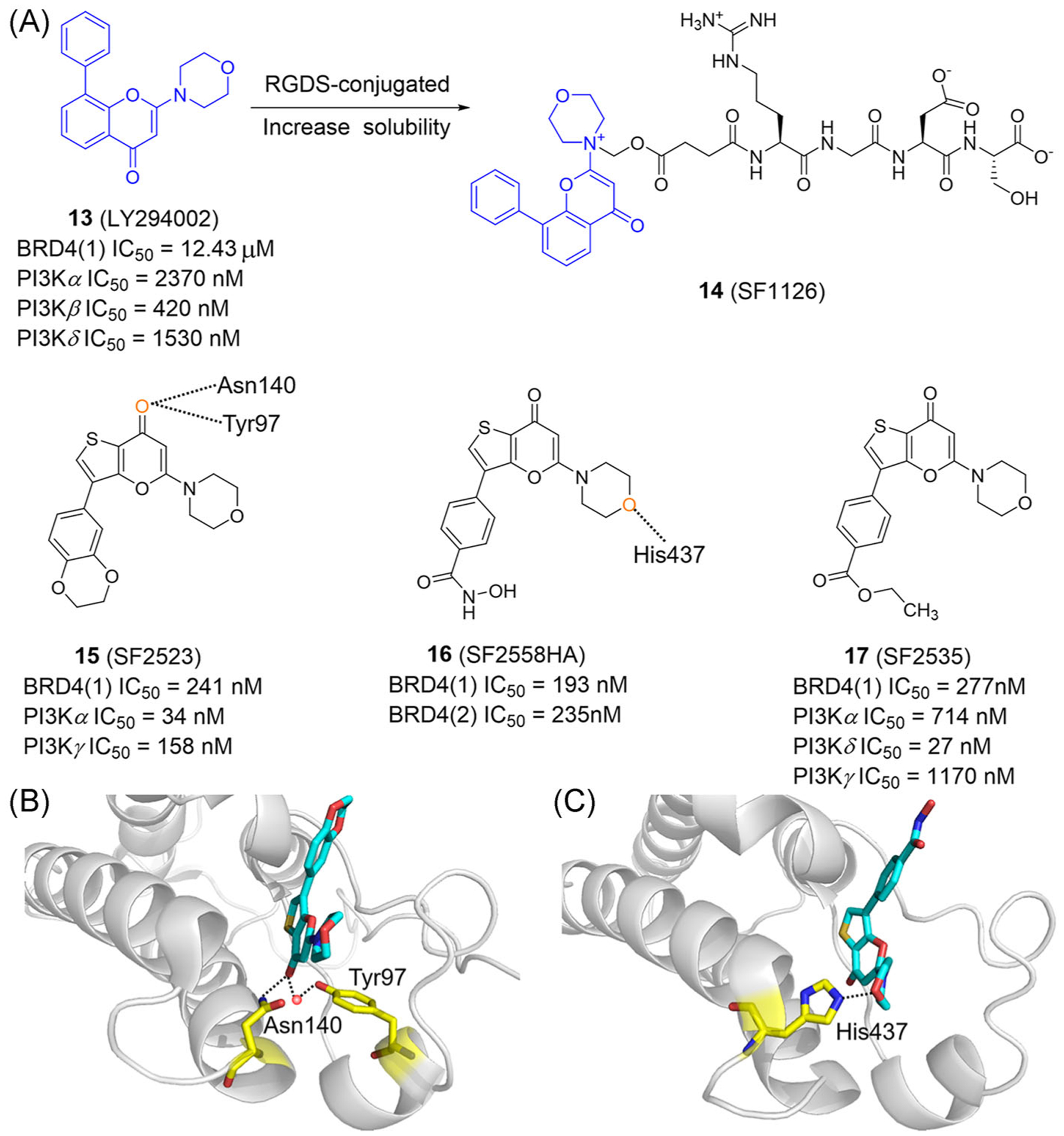
FIGURE 8.

Dual-target inhibitors of BET bromodomains and PI3K. (A) Chemical structures of dual inhibitors of BET bromodomains and PI3K. (B) X ray cocrystal structure of SF2523 bound to BRD4(1) (PDB ID: 5U28). (C) X ray cocrystal structure of SF2558HA bound to BRD4(2) (PDB ID: 5U2C). Protein is represented by gray ribbons, with compound-contacting residues indicated by yellow sticks. The compound is shown in sticks and colored according to the atom type (C, light blue; N, blue; O, red; S, yellow). Black dashed line = hydrogen bond; orange sphere = water molecule
