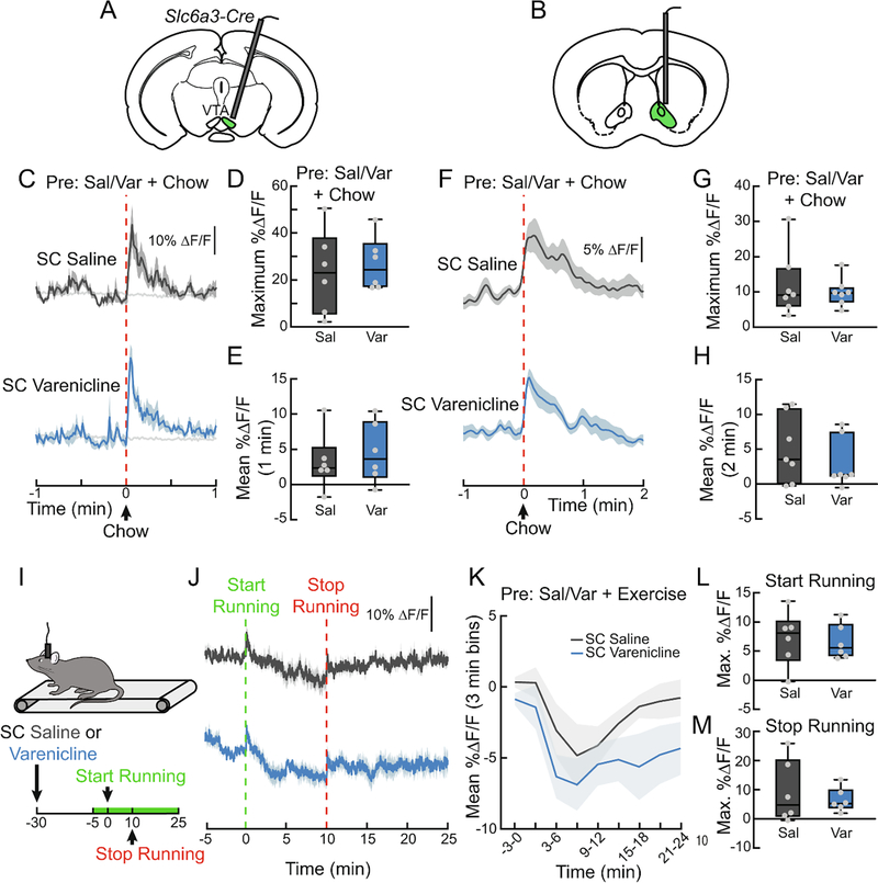
Figure 6. Varenicline does not affect DA responses to natural rewards.
(A) Schematic depicting GCaMP fiber photometry of VTA DAT neurons used in (C-E). (B) Schematic depicting GRAB-DA fiber photometry in the NAc of mice used in (F-H). (C) Average ΔF/F of VTA GCaMP6f signals of mice pretreated with SC injections of varenicline (1.5 mg/kg) or saline (0.9%) and then given a pellet of chow. Signals are aligned to chow presentation. Blue, 470 nm varenicline; dark grey, 470 nm saline; light grey, 450 nm. (D) Maximum ΔF/F of the 470 nm GCaMP6f signal shown in (C) (n=6/group, paired t-test). Blue, varenicline; grey, saline. (E) Mean ΔF/F of the 470 nm GCaMP6f signal shown in (C) (n=6/group, paired t-test). (F) Average ΔF/F of NAc GRAB-DA signals of mice pretreated with SC injections of varenicline or saline and then given a pellet of chow (n=7/group). (G) Maximum ΔF/F of the GRAB-DA signal shown in (F) (n=7/group, paired t-test). (H) Mean ΔF/F of the GRAB-DA signal shown in (F) (n=7/group, paired t-test). (I) Mice ran on a treadmill while their NAc DA signaling was monitored using fiber photometry. Mice were pretreated with SC varenicline or saline, ran for 10 min at 10 m/min, and then rested on the treadmill for another 15 min after running. Green on timeline represents duration of fiber photometry recording. (J) Average ΔF/F of GRAB-DA signals of mice from the experiment depicted in (I) (n=6/group). Signals are aligned to the start of running. (K) Mean ΔF/F (3 min bins) of GRAB-DA signals shown in (J) (n=6/group, two-way repeated measures ANOVA). (L) Maximum ΔF/F of the GRAB-DA signal shown in (J) during running (n=6/group, paired t-test). (M) Maximum ΔF/F of the GRAB-DA signal shown in (F) after running (n=6/group, paired t-test). Signals were aligned to the end of running. For photometry traces and line graphs, dark lines represent means and lighter, shaded areas represent SEM. For box and whisker plots, the box extends from the 25th to 75th percentiles, the line in the box represents the median, and the whiskers extend from the maximum to minimum values. Grey circles represent individual mice. See Table S1 for details on statistical analyses.

