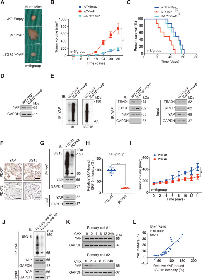
Fig. 3. YAP ISGylation is critical for LUAD malignant phenotype.
A–C Representative images (A), tumor volume (B), and survival analysis (C) for xenograft tumor formed by WT or ISG15−/− A549 cells with or without YAP overexpression. D YAP expression was analyzed by IB in xenograft tumors formed by WT or ISG15−/− A549 cells with YAP overexpression. E Co-IP experiments analyzing YAP ISGylation, ubiquitination, and interacted proteins in xenograft tumors formed by WT or ISG15−/− A549 cells with YAP overexpression. The YAP level in each co-IP sample was adjusted to the same protein content. F YAP and ISG15 expression in PDX#1 and PDX#2 were analyzed by IHC. Scale bar, 200 μm. G, H YAP ISGylation was analyzed by co-IP experiments and counted in PDX#1 and PDX#2 (n = 8). The YAP level in each co-IP sample was adjusted to the same protein content. I Tumor volume of PDX#1 or PDX#2 for indicated times. J YAP ISGylation was analyzed by co-IP experiments in Primary cell #1 and Primary cell #2. The YAP level in each co-IP sample was adjusted to the same protein content. K CHX (10 μg/ml) chase experiments were performed in Primary cell #1 and Primary cell #2. L Association between YAP half-life and relative YAP-bound ISG15 intensity was analyzed in 20 primary cell lines. The data are shown as the mean ± SD from three biological replicates (including IB). Data in B, I were analyzed using a two-way ANOVA test. Data in C were analyzed using a log-rank test. Data in H were analyzed using a student’s t test. Data in L were analyzed using a Spearman rank correlation analysis. **P < 0.01.

