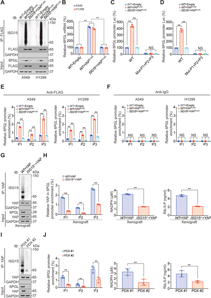
Fig. 6. YAP ISGylation stimulates 6PGL transcription.
A Co-IP experiments analyzing YAP ISGylation in WT or ISG−/− A549 and H1299 cells with or without overexpressing YAP, and YAP and 6PGL expression in Input sample were analyzed by IB. The FLAG level in YAPFLAG overexpressed co-IP samples was adjusted to the same protein content. B 6PGL mRNA level was measured in WT or ISG−/− A549 and H1299 cells with or without overexpressing YAP. C, D Luciferase activity was analyzed in WT or ISG15−/− A549 (C) and H1299 (D) cells co-expressing indicated 6PGL promoter–reporter with or without overexpressing YAP. E, F The enrichments of YAP at indicated regions of 6PGL promoter were calculated as the percentage of Input chromosomal DNA via ChIP in WT or ISG−/− A549 and H1299 cells with or without overexpressing YAP (E). Anti-IgG was used as parallel control (F). G Co-IP experiments analyzing YAP ISGylation in tumor xenografts overexpressing YAP with or without ISG15 knockout, and YAP and 6PGL expression in Input sample were analyzed by IB. The YAP level in each co-IP sample was adjusted to the same protein content. H The enrichments of YAP at indicated regions of 6PGL promoter, NADPH, and Rib-5-P level were analyzed in the same tumor xenografts as those in Panel G. I Co-IP experiments analyzing YAP ISGylation in PDX#1 and PDX#2, and YAP and 6PGL expression in Input sample were analyzed by IB. The YAP level in each co-IP sample was adjusted to the same protein content. J The enrichments of YAP at indicated regions of 6PGL promoter, NADPH, and Rib-5-P level were analyzed in PDX#1 and PDX#2. The data are shown as the mean ± SD from three biological replicates (including IB). Data in B–F were analyzed using a one-way ANOVA test. Data in H and J were analyzed using a student’s t test. **P < 0.01, NS nonsignificant.

