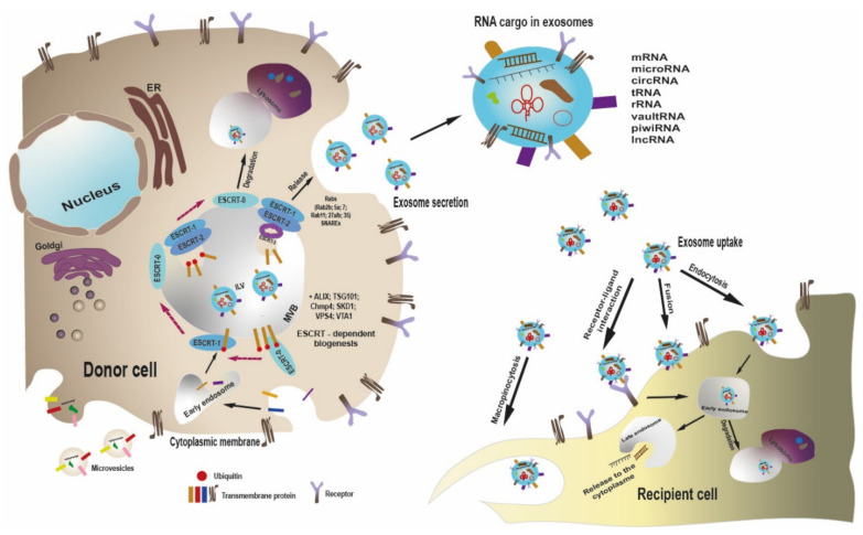
Figure 1.
Scheme of exosome biogenesis, release and uptake by recipient cell. Unlike microvesicles, which bud directly from the plasma membrane, exosomes are formed within the late endosome. When forming exosomes, ESCRT-0 binds the monoubiquitinated proteins, clusters them in specific patches on the endosomal membrane and passes to ESCRT-1. ESCRT-1 interacts with ubiquitinated proteins and activates ESCRT-2, which leads to formation of secondary invaginations of the late endosome and movement of ubiquitin-labeled proteins into ILV. ESCRT-2 recruits ESCRT-3, which exerts budding off of ILV, with MVB formation. MVB is subjected to either lysosomal degradation or plasma membrane fusion, with release of ILV as exosomes. A number of Rab GTPases and SNARE proteins are involved into exosome secretion. Exosomes contain a specific set of proteins and RNAs of several types. Exosomes can interact with recipient cell in several ways, such as receptor–ligand interaction, direct membrane fusion, macropinocytosis, or endocytosis. Within recipient cell, exosomes are either degraded or release their cargo to the cytosol, thereby modifying the recipient cell functions.

