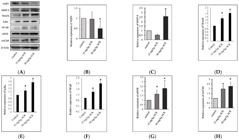
Figure 5.
Protein expression in the brain tissues of rats treated with 0, 15, or 30 mg/kg ACR for 28 days. Brain tissue lysates were used for the Western blot analysis. ACR inhibited AQP4 expression and induced MMP-9, TRAF6, IκBα, NFκB, nNOS, and nAChR expression (A). The quantitative data showing AQP4 (B), MMP-9 (C), TRAF6 (D), IκBα (E), NFκB (F), nNOS (G), and nAChR (H) protein levels. *: Significant difference versus the control group.

