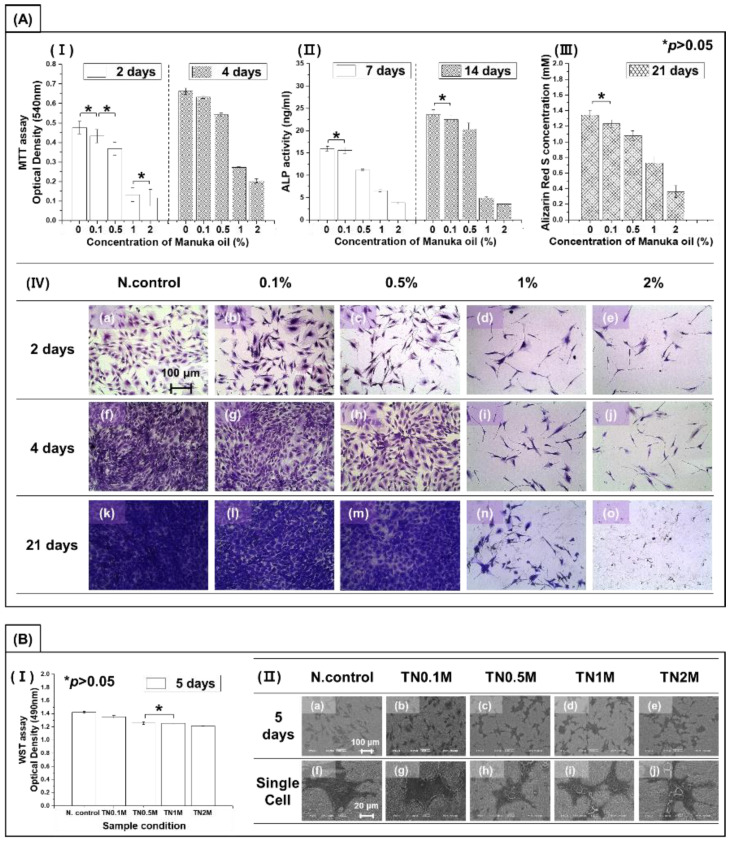
Figure 7.
(A) Cytotoxicity test of MC3T3-E1 osteoblast cells in the medium containing manuka oil: (I) proliferation (by MTT assay); (II) differentiation (by ALP assay); (III) quantification of calcium deposits (by ARS assay); and (IV) morphology of cells (by crystal violet staining). (B) Cell viability test on surfaces coated with different concentrations of manuka oil: (I) proliferation (by WST assay); and (II) morphology of cells (by osmium staining). * No significant difference (p > 0.05).

