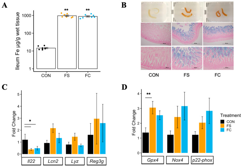
Figure 1.
Postnatal iron supplementation with ferrous sulfate (FS) or ferrous bis-glycinate (Ferrochel®; FC) leads to iron loading in the distal intestine and altered intestinal gene expression in pre-weanling rats. (A) Iron concentration in distal small intestine tissue (n = 7/group, 2 litters/group) collected from rat pups at PD 15 following 13 days of daily iron supplementation with FS or FC, or vehicle control (CON), assessed by atomic absorption spectrometry. Biological replicates are shown as individual data points with mean ± SEM. Differences in iron concentration among groups were detected with a Kruskal–Wallis test and Wilcox pairwise test. (B) Top row: distal small intestine samples exhibiting darkening effect of iron supplementation. Middle and bottom row: 10× (scale = 500 µm) and 20× (scale = 200 µm) objective microscope images of distal small intestine sections stained for iron with Perls’ Prussian blue staining (representative samples; n = 6/group, 3 litters/group). (C) mRNA expression of genes involved in regulation of mucosal immunity, quantified by real-time PCR (n = 5–8/group, 2 litters/group). (D) mRNA expression of genes involved in regulation of ferroptosis, an iron overload-dependent form of cell death. Gene expression values are shown as mean fold-change ± SEM. mRNA expression was normalized to Actb, and differences were detected among groups by one-way ANOVA and Tukey’s multiple comparisons. p-value summary: *, p < 0.05; **, p < 0.01.

