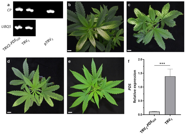
Figure 2.
A comparison of different TRV2-PDS vectors inoculated using vacuum infiltration. (a) The detection of the viral coat protein (CP) transcripts by RT-PCR in the cannabis line MF-219 infiltrated with pTRV2-PDS310 or pTRV2, and on pTRV2 plasmid as a positive control. UBQ5 was used as a control gene. Cannabis plantlets inoculated with pTRV1- and pTRV2-PDS310 (b), pTRV2-PDS486 (c), pTRV2-PDS424 (d), or pTRV2 (e) 15 days after vacuum infiltration (scale bar = 2 cm). (f) A quantitative real-time PCR analysis of PDS transcript levels in cannabis leaves infected with TRV2 and TRV2-PDS310. The data were normalized to UBQ5 with the standard error indicated by vertical lines. The significant difference between treatments (p ≤ 0.0006; n ≥ 3; ***) was calculated using Student’s t test.

