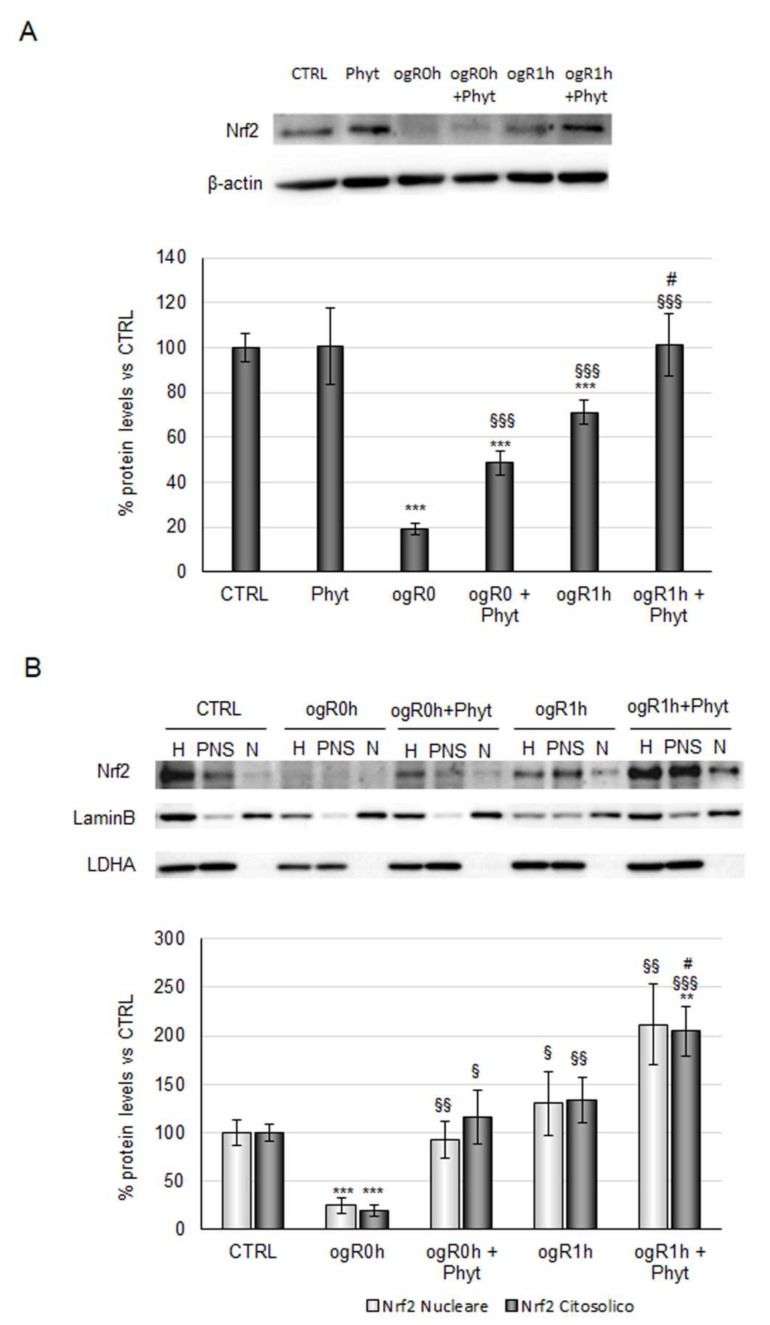
Figure 6.
Effect of coffee-pulp phytoextract on Nrf2 protein levels and cellular localization. (A) Cells pretreated or not for 24 h with 100 μg/mL Phyt and then subjected to OGD/ogR exposure were harvested in lysis buffer. Equal amounts of homogenate samples (as protein) were analyzed by SDS-PAGE electrophoresis and Western blotting. Nrf2 was detected with a specific antibody and revealed by enhanced chemiluminescence (ECL). Samples were normalized on β-actin immunoreactivity. (B) Cells pretreated or not for 24 h of 100 μg/mL Phyt and then subjected to OGD/ogR exposure were harvested in PBS solution and then subjected to fractionation protocol. Homogenate, nuclear, and cytoplasmic fractions of each sample were analyzed by SDS-PAGE electrophoresis and Western blotting. Nrf2, LaminB, and LDH were detected with specific antibodies and revealed by enhanced chemiluminescence (ECL). Nuclear Nrf2 and cytoplasmatic Nrf2 were normalized on LaminB and LDH immunoreactivity, respectively. Histograms, obtained from three distinct experiments, represent the percentage of protein levels with respect to control as mean ± S.E. ** p < 0.01, *** p < 0.001 versus CTRL (untreated cells); § p < 0.05, §§ p < 0.01, §§§ p < 0.001 versus ogR0h; # p < 0.05 versus ogR1h.

