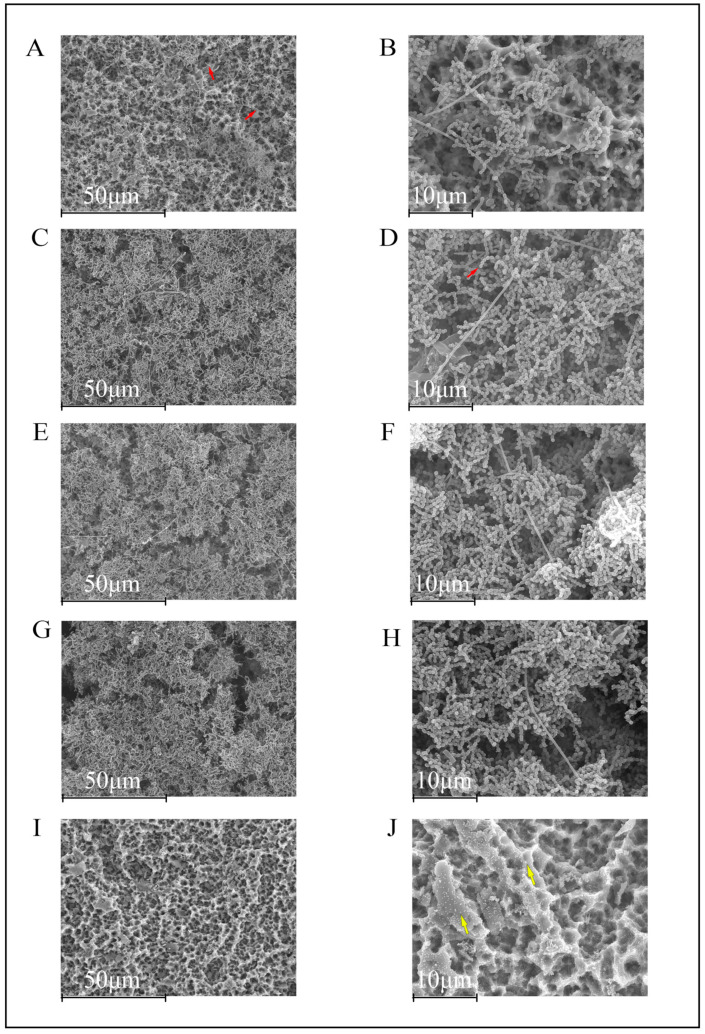
Figure 1.
Scanning electron microscopy images of 12 h biofilms on titanium discs (TiDs). F. nucleatum is recognized due to its morphological fusiform bacillus appearance. Bacteria colonized the entire rough surface of the phosphate buffer saline (PBS, negative control) coated TiDs (A,B). TiDs covered with undoped nanoparticles (NPs) (C,D), calcium nanoparticles (E,F), and zinc nanoparticles (G,H) presented a similar pattern of bacterial presence and distribution. The surfaces of TiDs covered with doxycycline nanoparticles (I,J) did not present biofilm formation. Bacilli and cocci are marked with red arrows. The NPs are sometimes visible (yellow arrows). Magnification: (A,C,E,G,I) 1000×; (B,D,F,H,J) 3000×.

