Fig. 2.
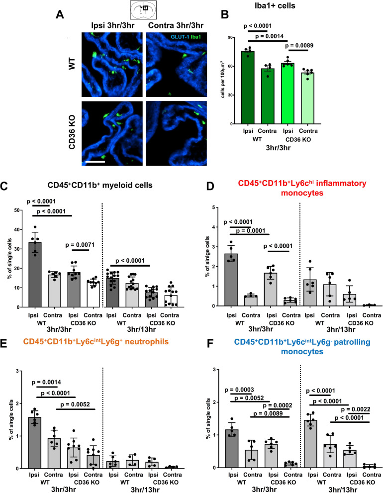
Accumulation of myeloid cells is attenuated in the ipsilateral CP of neonatal CD36 KO mice subjected to tMCAO. A Representative images of CPs at the level of the lateral ventricles immunostained with GLUT-1 (blue) and Iba1 (green) ipsilateral and contralateral to tMCAO in WT and CD36 KO mice at 3 h reperfusion. Scale bar = 50 µm. B Quantification of the number of Iba1+ cells in the ipsilateral and contralateral CPs in WT and CD36 KO mice at 3 h reperfusion. C–F Quantification of CD45+CD11b+ myeloid cells (C), CD45+CD11b+Ly6chi inflammatory monocytes (D), CD45+11b+Ly6cintLy6g+ neutrophils (E), and CD45+11b+Ly6cintLy6g− patrolling monocytes (F) ipsilateral and contralateral to tMCAO in WT and CD36 KO mice at 3 h and 13 h reperfusion. Two-way ANOVA with post-hoc Tukey’s Multiple Comparison test was performed to compare groups with multiple independent variables (B–F). Dots represent individual mice. Data are shown as mean ± SD. Individual p values are listed within figures for data that are significant

