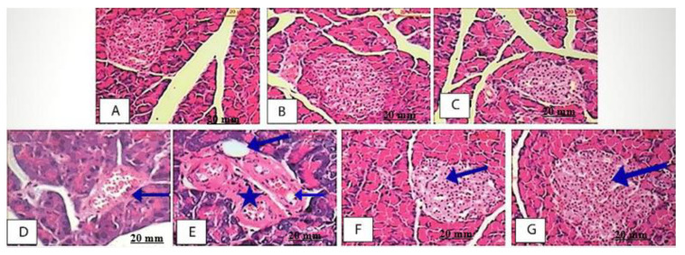
Figure 9.
The histological investigation of pancreas. (A–C): Pancreas sections of NC, AE500-NC and AE 250-NC groups respectively, showing dense staining acinar cells and a light-staining islet of Langerhans; (D): Diabetic rat showing the acinar cells around the islets though seemed to be in normal proportion with up normal morphology with shrunken islet is and intra islet hemorrhage (blue arrow). (E): Diabetic rat showing degenerative islet of Langerhans (asterisk) with different vacuoles size (long arrow) and hemorrhage (short arrow); (F,G): AE 500-DC rat showing the exocrine pancreas appearing.

