
Figure 1.
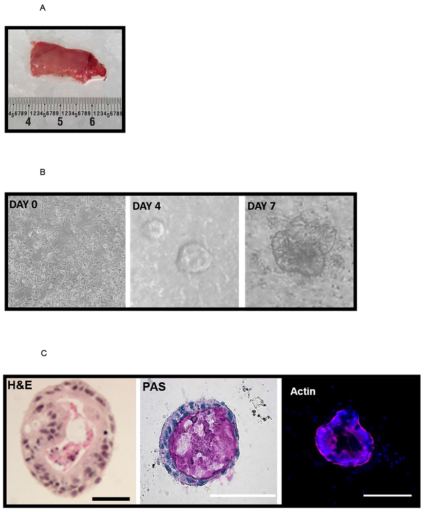
[A] Discarded intestinal segments are collected at the time of surgery. A piece of neonatal ileum is shown. The anatomic location is noted, and the specimen is immediately placed on ice. Samples are processed as described in the methods.
[B] Crypt isolation and culture leads to generation of human enteroids. On day 0 a crypt culture shows characteristic cells with numerous rod-shaped cells, and by day 4 sphere-like enteroids can be seen. The day 7 enteroid is well developed with budding features. Experiments were repeated in triplicate with enteroids generated from 4 different samples.
[C] Structural appearance of the enteroids. The histological appearance is demonstrated by staining with hematoxylin and eosin. A central lumen is seen, encompassed by a polarized exterior. The nuclei are dark and the cytosol is pink. Goblet cells are present and staining with PAS reveals mucin (pink) within the center of the enterocyte. Experiments were repeated in triplicate with enteroids generated from four different samples. Immunofluorescence reveals the actin cytoskeleton (pink) and nuclei (blue + DAPI). Scale bar 50 μm.