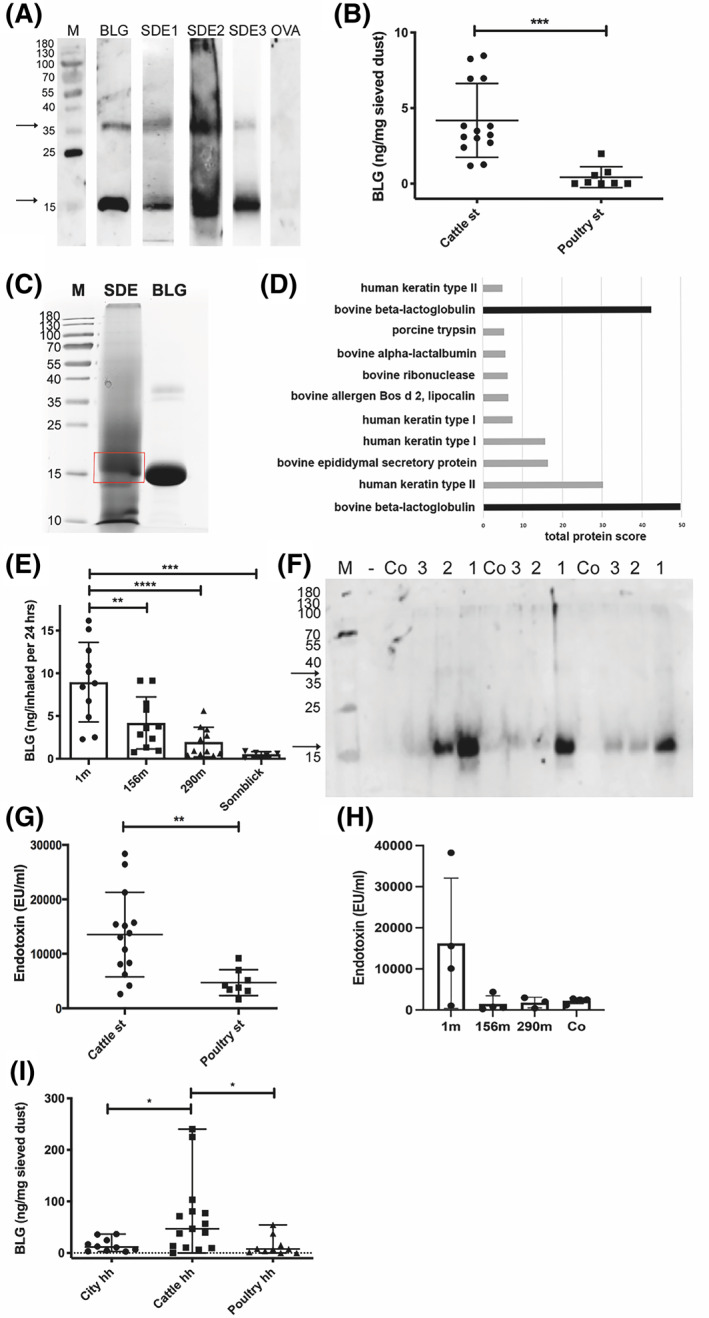
FIGURE 1.

BLG and endotoxin levels in stable dust and ambient air of cattle farms. (A) BLG in dust of cattle stables, which was collected by different methods, detected in immunoblot: stable dust extract SDE1 (Table S1, set 1, Vet) = dust wiped from elevated surfaces; SDE2 (Table S1, Set 1, Bav) = dust deposition on cardboard box over 3 weeks; SDE3 (Table S1, set 3, Vet) = dust collected by air filtering (1 representative example of at least 3 repetitions per collection method is shown; due to time interval between examination, strips of different individual blots are shown). (B) Stable dust extracts (SDE), all collected by wiping, from cattle farms (n = 14; Table S1, set 2, C1–14) and poultry stable (n = 8; Table S1, set 2, P1‐8) investigated by BLG‐specific ELISA (mean +/‐ SD; representative of 3 repetitions). (C) BLG in stable dust (sample SDE 2) confirmed by MS/MS‐LC in SDE separated via SDS‐PAGE, stained by Roti‐Blue® and the major band around 18 kDa excised. (D) Protein of the excised band in MS/MS‐LC (proteins UniProtKB P02754 and B5B0D4 with difference of 2 amino acids in sequence). (E) BLG‐concentration in air samples at different distances from cattle stable (Table S1, set 4, n = 4 filter/distance), extrapolated to the human respiratory volume per 24 h, determined in ELISA (1 m = outside the stable in front of open window; 156 m and 290 m distance from cattle stable, sampled on cellulose filters; at the mountain site Sonnblick at 3106 m above sea level, sampled on quartz fiber filters); and (F) in immunoblot with bovine BLG‐specific antibodies (1 = 1 m, 2 = 156 m, 3 = 290 m, Co = empty control filter; 3 different time points from E shown). (G) Levels of endotoxin were determined in dust samples of cattle (n = 14) and poultry stable (n = 8) (Table S1, set 2) by LAL test, and (H) in dust samples collected in different distances to cattle stable. (I) Occurrence of BLG in different households (hh). BLG in sieved bed dust samples from beds of cattle farm households (Cattle hh. N = 14) , poultry farm households (Poultry hh, n = 8) or urban apartments (Urban hh, n = 10), detected by an anti‐BLG antibody in ELISA (mean of 2 repetitions). BLG (commercial beta‐lactoglobulin) = positive control; OVA (ovalbumin) and Co (empty control paper filter) = negative control. M: protein weight marker in kDa. Arrows indicate monomeric (around 18 kDa) and dimeric (38 kDa) BLG. *p < 0.05, **p < 0.01, ***p < 0.001, ****p < 0.0001
