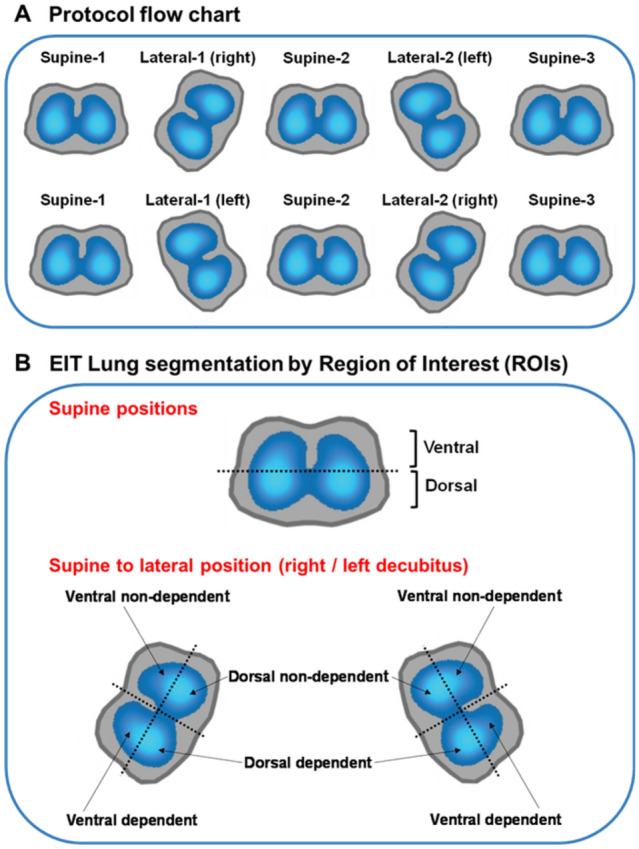
Fig. 1.

Protocol flowchart and EIT lung segmentation. The protocol flowchart (A) is shown at the top. The positioning sequence begins with the less ventilated lung evaluated by EIT positioned upwards in L1. In section B is shown the EIT lung segmentation by ROIs. To compare changes during supine position, the lungs were segmented into two equally sized ROIs: ventral and dorsal. To compare changes from supine to lateral position, the lungs positioned upwards (non-dependent) or downwards (dependent lung) during L1 or L2, were segmented into four ROIs or quadrants: ventral non-dependent, dorsal non-dependent, dorsal dependent, and ventral dependent. EIT: electrical impedance tomography; ROI: region of interest; L1: first lateral; L2: second lateral
