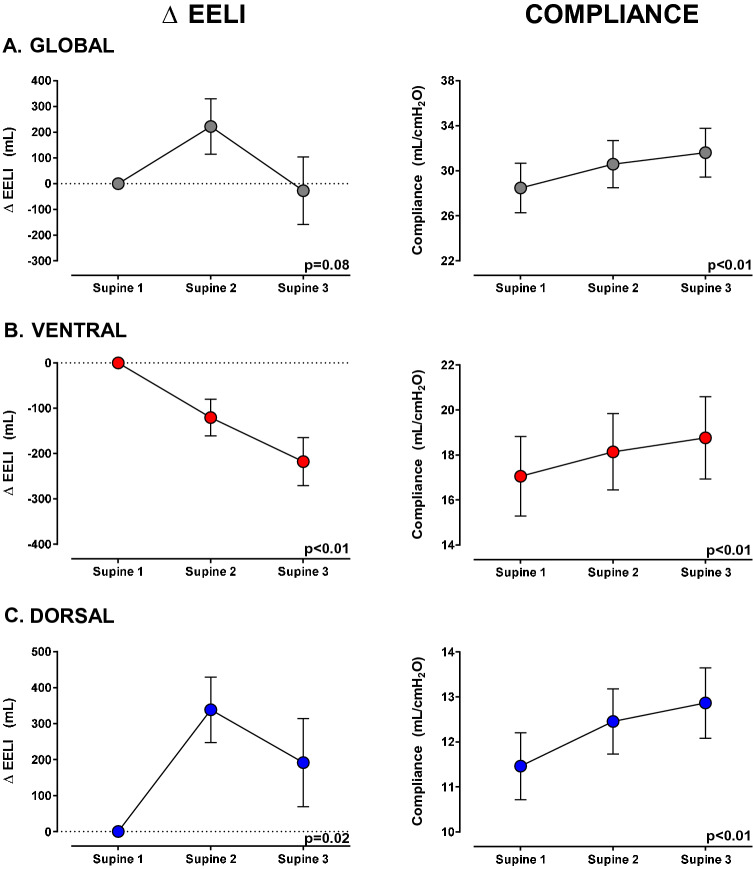
Fig. 2.
End-expiratory lung impedance and compliance changes during supine position steps, from supine-1 (baseline) to supine-3 (after second lateral positioning), segmenting the lung into ventral and dorsal areas. The left column shows changes in the EELI, and the right column shows changes in compliance. The figures on the upper panel A show global lung changes, while the figures in the middle panel B show changes in the ventral (anterior half) part of the lung, and the figures on the bottom panel C show changes in the dorsal (posterior half) of the lung. The left column shows that global EELI did not change between supine-1 and supine-3, but the EELI decreased in the ventral region and increased in the dorsal region. This redistribution of EELI was accompanied by a progressive increase of the global and regional compliance (both ventral and dorsal) (right column). Δ EELI (mL): end-expiratory lung impedance change. Data are shown as mean ± SEM (standard error of mean). Mixed model was used for statistical analysis

