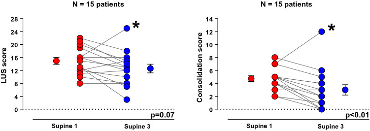
Fig. 3.
Evaluation of aeration by lung ultrasound between supine-1 (baseline) and supine-3 (after second lateral positioning). The analysis of the individual data shows an improvement in LUS score (left figure) and consolidation score (right figure) in the majority of patients. A non-responder patient is identified with asterisk *LUS score: lung ultrasound score. Paired t test was used for the analysis of LUS score and Wilcoxon signed-rank test for the consolidation score

