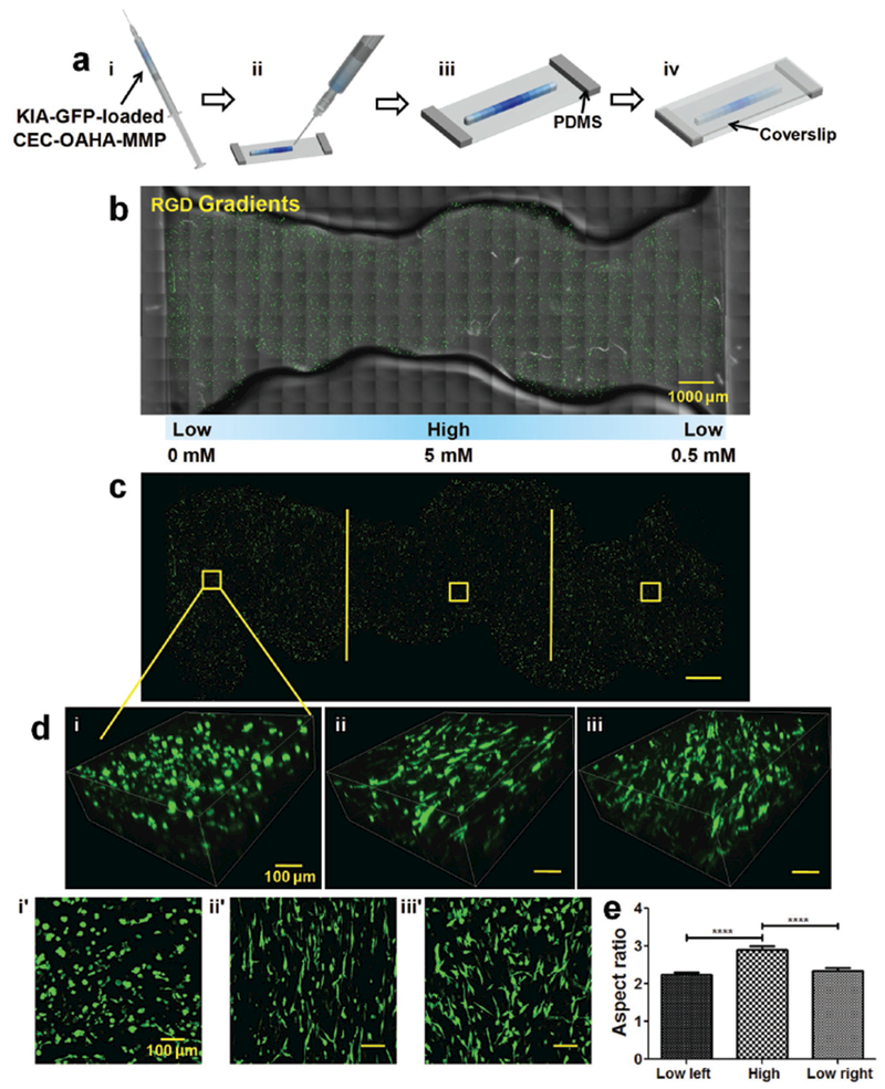
Figure 3.

Generation of cell-loaded gradient hydrogel constructs and cancer cell response to RGD gradients a) Preparation of KIA-GFP-loaded CEC-OAHA-MMP modular gradient hydrogel constructs for microscopic observation. (i) The tiny discs of the CEC-OAHA-MMP hydrogels with various concentrations (here illustrated with blue color) were loaded into the syringe and then injected to generate stripe-shaped construct onto a glass slide equipped with 2 mm thick PDMS (ii and iii). (iv) A coverslip was placed over the injected hydrogel to obtain a 2 mm thick hydrogel stripe. b,c) Confocal images of the entire injected KIA-GFP-loaded CEC-OAHA-MMP hydrogel stripe-shaped construct with RGD gradient distributions from 0 to 5 × 10⁻³ to 0.5 × 10⁻³ m after culturing for 3 d (the encapsulated KIA-GFP cells are green), scale bar: 1000 μm. d) 3D and z-axis maximum projection views of confocal images of encapsulated KIA-GFP cell spatial distribution and morphology from the left lower RGD section (i and i′), the high RGD section (ii and ii′), and the right lower RGD section (iii and iii′), scale bar: 100 μm. e) Aspect ratio analysis of the encapsulated KIA-GFP cells from different gradient sections (for each section, we randomly selected ten views for the calculation). Significance levels were set at ****P < 0.0001.
