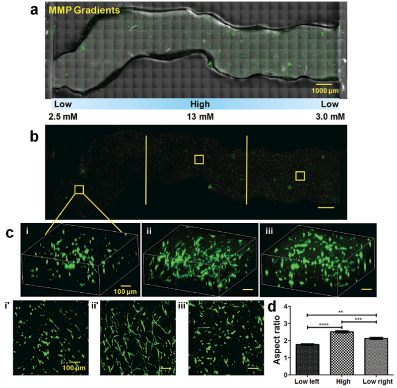
Figure 4.

Cancer cell response to MMP-sensitive cross-linker gradients. a) Confocal images of the entire injected KIA-GFP-loaded CEC-OAHA-MMP hydrogel stripe-shaped construct with MMP-sensitive cross-linker gradient distributions from 2.5 × 10⁻³ to 13 × 10−3 to 3.0 × 10⁻³ m after culturing for 3 d (the encapsulated KIA-GFP cells are green), scale bar: 1000 μm. b) Confocal images of the entire view of encapsulated KIA-GFP cells, scale bar: 1000 μm. d) 3D and z-axis maximum projection views of the confocal images of the encapsulated KIA-GFP cell spatial distribution and morphology from the left lower MMP-sensitive cross-linker section (i and i′), the high MMP-sensitive cross-linker section (ii and ii′), and the right lower MMP-sensitive cross-linker section (iii and iii′), scale bar: 100 μm. e) Aspect ratio analysis of the encapsulated KIA-GFP cells from different gradient sections (for each section, we randomly selected ten views for the calculation). Significance levels were set at **P < 0.01, ***P < 0.001, and ****P < 0.0001.
