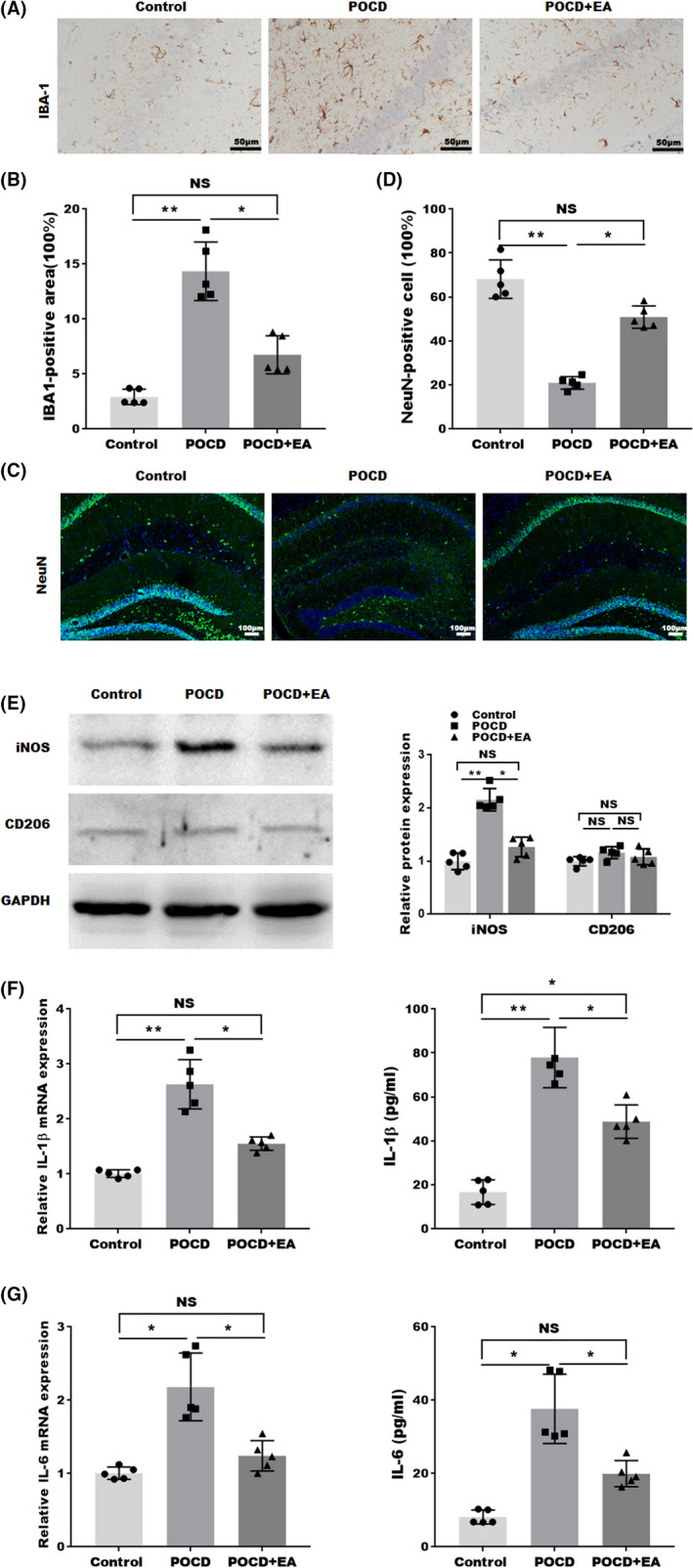
FIGURE 2.

EA treatment decreased neuroinflammation in the hippocampus of aged POCD mice. (A) Representative images of Iba1 staining and (B) quantification of Iba1‐positive area in hippocampus. (C) Representative images (NeuN: green; DAPI: blue) and (D) quantifications of nucleus staining in the hippocampus. (E) Western blotting and semiquantifications for iNOS and CD206 protein level in hippocampal tissues. (F) The mRNA and protein expression IL‐1β in the hippocampal tissue detected by qPCR and ELISA from hippocampal tissues. (G) The mRNA and protein expression IL‐6 in the hippocampal tissue were detected by qPCR and ELISA from hippocampal tissues. Each data point was mean ± SD (n = 5). *p < 0.05, **p < 0.01
