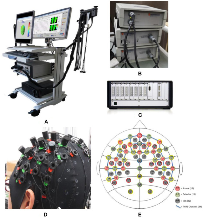
Figure 2.
Acquisition equipment and measuring cap. (A) View of the acquisition equipment; (B) Acquisition amplifier of EEG; (C) Laser light source of fNIRS; (D) View of measuring cap; and (E) Channel location of EEG and fNIRS. Where the red circles represent the 16 light sources of fNIRS, the green circles are 15 detesctors of fNIRS, the blue lines are 44 channels of fNIRS, and the gray circles represent 32 EEG electrodes.

