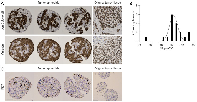
Figure 3.
IHC staining of breast cancer spheroids reveals the distribution of fibroblasts and epithelial cells within the spheroids. (A) Three tumor spheroid replicates and the original tissue from one patient sample (patient ID #31) were stained by IHC for epithelial (panCK) and fibroblast (vimentin). (B) Histogram analysis of panCK quantification shows that the majority of spheroids contains around 40–45% epithelial cells (n=18). (C) Three tumor spheroid replicates and HDF-only spheroids were stained by IHC for Ki-67. Positive Ki-67 staining is only observed in the tumor spheroids. Scale bar: 100 µm. IHC, immunohistochemistry; panCK, pan cytokeratin; HDF, human dermal fibroblasts.

