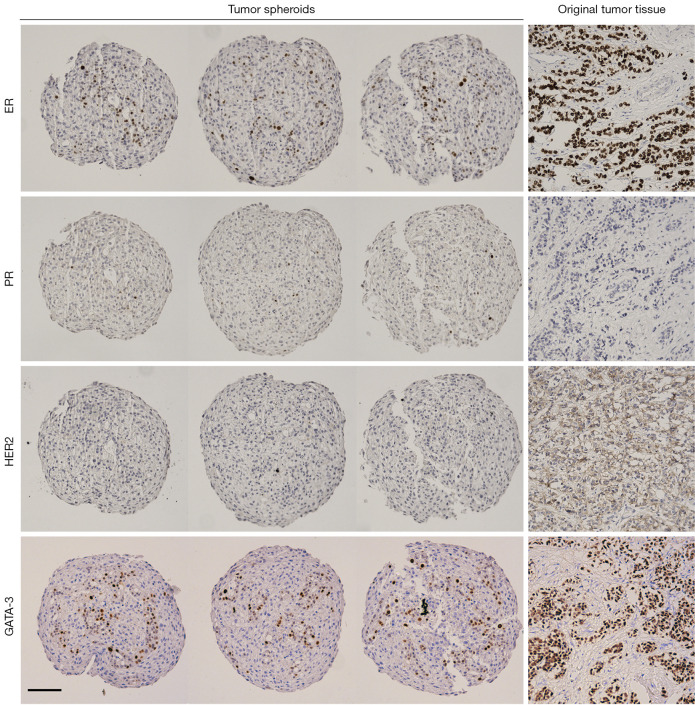
Figure 4.
IHC staining of breast cancer spheroids reveals the heterogenous mixture of cellular components within the spheroids. Three tumor spheroid replicates and original tissue from one patient sample (patient ID #31) were stained by IHC for breast cancer markers (ER, PR, HER2 and GATA-3). The IHC staining of the spheroids resembles that of the original tissue (ER+, PR−, HER2−; luminal B1). Scale bar: 100 µm. IHC, immunohistochemistry.

