Fig. 2.
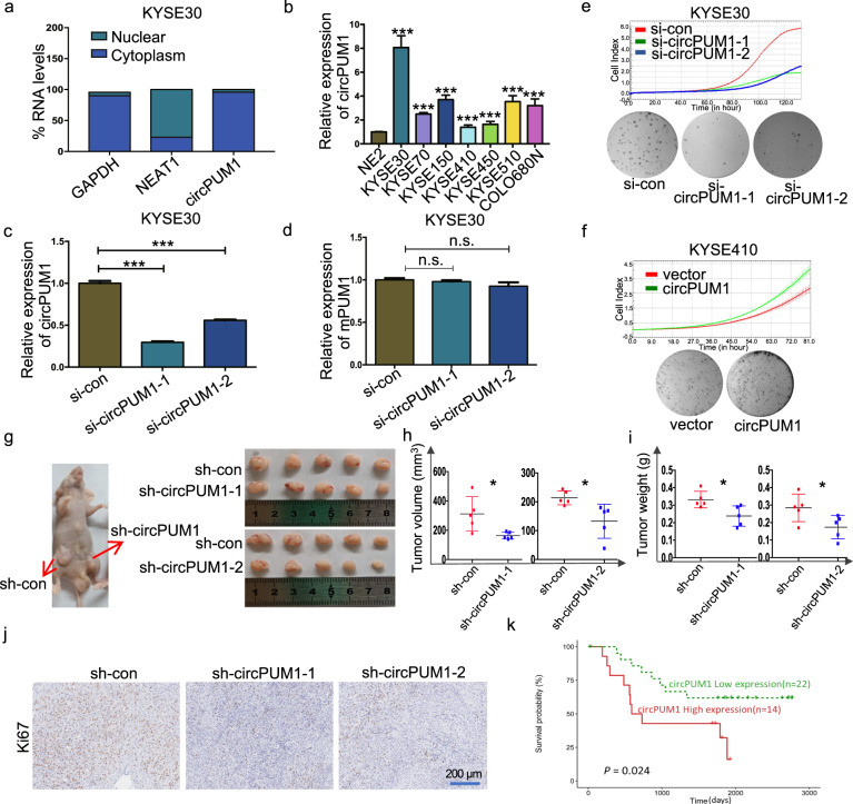
CircPUM1 enhances the tumorigenicity of ESCC cells in vitro and in vivo. a Fractionation of KYSE30 cells following by RT-qPCR was used to determine the localization of circPUM1. GAPDH served as the cytoplasmic expression control and NEAT1 severed as the nuclear expression control. b RT-qPCR analysis of circPUM1 expression profile in ESCC cell lines that normalized by normal esophageal epithelial cell line NE2. c RT-qPCR analysis of circPUM1 after knocking down circPUM1 in KYSE30 cell. d RT-qPCR analysis of mPUM1 after knocking down circPUM1 in KYSE30 cell. e The growth curve that monitored by the RTCA-MP system and colony formation analyses following the depletion of circPUM1 in KYSE30 cell. f The growth curve that monitored by the RTCA-MP system and colony formation analyses following the overexpression of circPUM1 in KYSE410 cell. g Macroscopic appearance of the xenograft tumors in nude mice after injection with KYSE30-sh-con (left side on both), KYSE30-sh-circPUM1-1 (right side) (up) and KYSE30-sh-circPUM1-2 (right side) (down). Xenograft tumors were isolated, KYSE30-sh-con (up on both side), KYSE30-sh-circPUM1-1 and KYSE30-sh-circPUM1-2 (down on both side). h–j Comparison of the growth of KYSE30 xenografts expressing sh-con versus sh-circPUM1 in tumor volume (h) and tumor weight (i); IHC that with Ki67 antibody staining (j), the scale bar is 200 μm. k Elevation of circPUM1 in ESCC patients was associated with worse overall survival of ESCC patients

