Figure 1 .

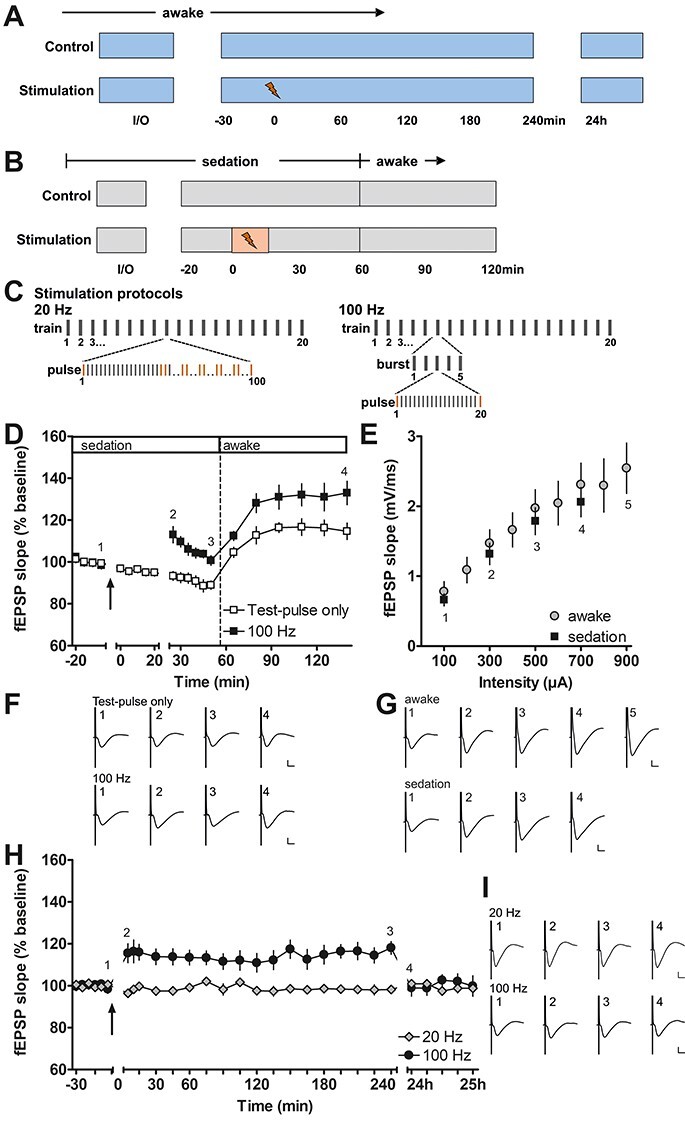
Long-term potentiation in the piriform cortex induced by prolonged electrical stimulation of the olfactory bulb in sedated and awake behaving rats. (A) Experimental procedure for testing prolonged patterned stimulation of the olfactory bulb, while recording in the anterior piriform cortex in awake behaving animals. During the control experiment, the stability of the evoked responses was confirmed. Only animals exhibiting stable evoked responses over the 25 h monitoring period were tested with prolonged patterned stimulation (20 and 100 Hz protocols). (B) Experimental procedure during fMRI-like conditions under medetomidine sedation. The total duration of medetomidine sedation was similar during control and stimulation (100 Hz protocol) experiments. (C) Scheme depicting the patterns of the 20 Hz (left) and 100 Hz (right) stimulation protocols. Both protocols consist of 20 trains with 100 pulses applied during each train. In the 20 Hz protocol, the 100 pulses of each train are applied consecutively at 20 Hz. Ten evoked responses to pulses 1, 20, 21, 40, 41, 60, 61, 80, 81, and 100 (marked in orange) were analyzed for each train during 20 Hz stimulation in behaving rats. In the 100 Hz protocol, each train contains five bursts and each burst contains 20 pulses applied at 100 Hz. For the analysis during 100 Hz stimulation, the responses (marked in orange) evoked by the 1st and 20th pulse of each burst were analyzed. (D, F) Patterned stimulation of the olfactory bulb at 100 Hz applied under medetomidine sedation (n = 7, black squares) induces long-term potentiation (LTP) lasting at least 2 h in the anterior piriform cortex. (Arrow indicates the start of 100 Hz stimulation). Where only test pulses were applied to the olfactory bulb (white squares), evoked potentials recorded in the piriform cortex decrease in magnitude during sedation and increase after the animals are awake. (F) Representative evoked responses recorded (1) 5 min before, (2) 5 min after, (3) 30 min after, and (4) 2 h after the end of patterned stimulation. (E, G) Comparison of input–output relationship for fEPSP responses recorded in the piriform cortex of sedated (n = 7) and awake (n = 9) rats evoked by test pulses applied to the olfactory bulb. All values shown in these graphs were recorded at the start of the experiments where the 100 Hz protocol was applied. (G) Representative evoked responses recorded during the evaluation of the input–output relationship at a stimulus intensity of (1) 100 μA, (2) 300 μA, (3) 500 μA, (4) 700 μA, and additionally in awake animals (5) 900 μA. (H, I) Patterned stimulation at 100 Hz, applied to the olfactory bulb in awake behaving rats (n = 9), induces LTP lasting for at least 4 h in the anterior piriform cortex. In contrast, 20 Hz stimulation (n = 8) does not change synaptic strength. Arrow indicates application time of patterned stimulation at 20 Hz, or 100 Hz. Graphs comparing 20 and 100 Hz stimulation to test–pulse stimulation are provided in Supplementary Fig. 2. (I) Representative evoked responses recorded (1) 5 min before, (2) 5 min after, (3) 4 h after, and (4) 24 h after patterned stimulation. F, G, I calibration: vertical bar: 1 mV, horizontal bar: 5 ms. D, E, H mean ± SEM.
