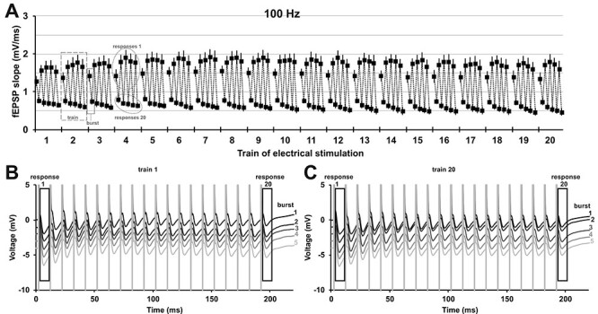
Figure 2 .

Patterned stimulation of the olfactory bulb changes synaptic responses in the anterior piriform cortex in a distinct manner. (A) Analysis of every 1st and 20th evoked response of each burst during patterned stimulation at 100 Hz (n = 7) revealed that the fEPSP changes under medetomidine sedation (mean ± SEM is shown). The 100 Hz stimulation consisted of 20 trains, each train contained five bursts, and every burst consisted of 20 pulses applied at 100 Hz (see Fig. 1C). Evoked responses of pulse 1 and pulse 20 of every burst are displayed (the two analyzed responses of burst 1 in train 3 are marked with a box with a solid line). Thus, for every train (train 2 is marked as example with a box with a dashed line), a total of 10 responses were analyzed (five 1st responses and five 20th responses, example in circle with dotted outline). (B, C) Representative examples of evoked responses of train 1 and train 20 recorded during prolonged patterned stimulation. Every 1st (response 1) and every 20th (response 20) evoked response of each burst was analyzed for (A) and are marked with an outline. The trace of each subsequent burst is plotted with an offset of −1 mV to the previous burst. (B) Evoked responses of train 1 during each of the five bursts of patterned stimulation (burst 1–5: top to bottom, black to light gray). (C) Evoked responses of train 20 during each of five bursts of stimulation (burst 1–5: top to bottom, black to light gray).
